
Scientific Artwork
|
Matthew Clark
Art of the Science: Neural Modeling
3D imaging shows how molecule shape affects activity
What are we looking at?
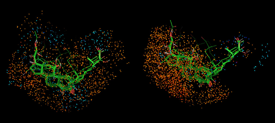
The images are a set of steroids, depicted inside fields computed via a neural network model that shows where the atoms contribute most to their bioactivities. When atoms are near the bluer regions, they make the molecule more active. The image maps where the shape and the electric field generated by the atoms contribute to the biological activity.
How was the image generated?
The image was generated by fitting computed 3D fields of the aligned molecules using a neural net “AI” model created with Google TensorFlow. The neural network training learns which atoms are most impactful on the molecule’s biological activity. The images were then rendered with the open-source tool Pymol.
What are researchers learning from this image?
Researchers use this Comparative Molecular Field Analysis (CoMFA) model to predict the biological activity of molecules and understand how the shape affects activity. This helps medicinal chemists design more active compounds.
Can this help improve the pace of drug development?
This technique improves drug development by allowing scientists to use the colored field maps to design more active compounds and test their activity with the predictive model before synthesizing the molecules. While there are many methods to predict the effect of molecules, this method is unique because it provides a visual guide for how to change a molecule’s 3D structure to improve activity.
Tell me something cool about yourself.
I am also a watchmaker and watch designer, not designing the decorative cases but the internal mechanisms of mechanical watches, specializing in high-accuracy timepieces.
Matthew Clark is the Global Head of Science Analytics at Charles River Laboratories, and earned his PhD in Chemistry from the University of Alabama.