

Cell & Gene Therapy
|
Anne Lodge, PhD
Ask A Scientist: What’s the Difference Between M1 and M2 Macrophages?
Macrophages are a diverse group of white blood cells known for eliminating pathogens through phagocytosis. In the past, macrophages were classified by the organ in which they were found: Kuppfer cells in the liver, Langerhans cells in the skin, microglia in the brain and spinal cord, osteoclasts in the bone.
Scientists continue to debate whether macrophages originate in each organ or migrate there from the bone marrow. To clear up some of this debate, the current taxonomy has shifted away from organ-specific macrophages to M1 and M2 macrophages.
Defining M1 and M2 Macrophages
This classification is based upon macrophage polarization rather than macrophage location.
M1 macrophages are classically activated, typically by IFN-γ or lipopolysaccharide (LPS), and produce proinflammatory cytokines, phagocytize microbes, and initiate an immune response. M1 macrophages produce nitric oxide (NO) or reactive oxygen intermediates (ROI) to protect against bacteria and viruses.
M2 macrophages are alternatively activated by exposure to certain cytokines such as IL-4, IL-10, or IL-13. M2 macrophages will produce either polyamines to induce proliferation or proline to induce collagen production. These macrophages are associated with wound healing and tissue repair.
There are three types of M2 macrophages: M2a, M2b, and M2c.
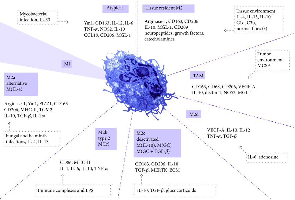
Source: Tamás Rőszer, “Understanding the Mysterious M2 Macrophage through Activation Markers and Effector Mechanisms,” Mediators of Inflammation, vol. 2015, Article ID 816460, 16 pages, 2015. doi:10.1155/2015/816460
M2 macrophages also contribute to the formation of extracellular matrix and do not produce nitric oxide or present antigen to T cells. Tumor-infiltrating macrophages are typically classified as M2, although some classify them as myeloid-derived suppressor cells (MDSC).

Human Immune and Stem Cells Product Catalog
Charles River is a trusted provider of research-use and GMP-compliant human immune cells and stem cells for advanced therapies and basic research. Our portfolio also includes disease state biospecimens for disease-focused research.
Download Catalog
Differences Between M1 and M2 Macrophages
| M1 Macrophages | M2 Macrophages | |
|---|---|---|
| Activation | Classical (Th2) | Alternative (Th2) |
| Stimuli | IFN-y, LPS | IL-4, IL-13, Immune Complexes, LPS, Glucocorticoids, etc. |
| Proinflammatory Cytokines | High Levels | Low Levels |
| Antigen Presentation | Yes | No |
| Nitric Oxide Production | Yes | No |
| Function | Kill Microbes | Build Extracellular Matrix |