

Biologics
|
Ulrike Herbrand, PhD
CAR-T Cell Therapy for Solid Tumors: Biological Activity Testing for Lot Release
The determination of biological activity of CAR-T cells plays a key role in defining the quality of the product
NOTE: This article is also available in another language (한국어).
T cell therapies are novel and, in some cases, curative, but they also present some formidable limitations. Follow our three-part series, Driving the CAR-T, and see how a CAR-T cell therapy is engineered and produced, and how it is tested in vivo. Today we are looking how to assess the effects in vitro to determine the biological activity.
Despite progress in recent years, finding a mechanism of action (MoA) reflecting cell-based bioactivity assay for complex therapeutics is still a challenge. The challenge is even bigger if the complexity of the drug, such as a CAR-T drug, is higher than that of the MoA reflecting assay itself.
The biological activity of CAR-T cells is impacted by many different factors including donor variability, transfection efficiency, CAR structure, gene vectors, cell culture conditions and cell proportions. By knowing all these sources of variability, the MoA reflecting bioassay should be capable of measuring the relative biological functionality compared to a reference batch. And if you can accomplish, it will go a long way toward ensuring quality and consistency in the manufacturing process. You should also be able to determine stability and comparability when production processes change. Given all these challenges a lot release method needs to be reliable, reproducible, suitable for standardization and easy to run in the lab if you want to overcome issues related to the product variability.
The biological activity is highly individual for each CAR-T product and is dependent on the target. Therefore the “right” MoA reflecting bioassays needs to be chosen carefully. Each individual product requires establishment of an individual assay because dependent on the target a suitable target cell line needs to be established and all assay conditions including effector-to-target ratio and incubation times need to be optimized. The suitability of whatever biological activity method is chosen will be evaluated by regulatory agencies on a case-by-case basis. In the best-case scenario, the MoA reflecting assay is based on a biologic characterization that confirms the relevant biologic function.
MoA-reflecting biological activity readouts
Potential readouts for bioactivity testing are either based on release of cytokines upon activation of the CAR-T cells in the present of target cells, such as IFNg or TNF or activation of target-specific killing of target cells by CAR-T cells or activation of cell surface markers, such as CD25 or CD95. Here are biological activity assays that come into play:
-
Cytokine release: Cytokine release assays are traditionally still measured by ELISA, even though they bring background issues, limited dynamic range and lots of handling steps that come from adding the different reagents and wash in between. These downsides can be overcome, however, by choosing newer wash-free labelling technologies that are superior to classical ELISA assays (see illustration (Figure 1) below). The luminescence readout is highly sensitive, allows for an improved dynamic range while the wash-free approach is faster and less susceptible to lab errors. The disadvantage of cytokine release assays is that it can be quite difficult to determine if target-induced cytokine secretion, while certainly an aspect of T-cell activation, is truly MoA-reflective of CAR-T activity. Showing the death of the target cells in the presence of activated CAR-T cells is probably a better option.
-
Cell killing: A cell-killing assay, even though highly reflective of the MoA is even more tedious and tends to have significant issues with non-specific background. This can be overcome with specific genetically modified target cells that allow for a stream-lined life-cell analyses with luminescence readout and great dynamic range (see Figure 2 below).
-
Activation of cell surface markers: Classical flow cytometry to measure the activation of certain markers has limited throughput and requires experienced staff to deal with the complex data generated by co-cultivated CAR-T effector cells and specific target cells.

Figure 1: Promega LUMIT Technology
The LUMIT Technology is a homogenous wash-free and fast alternative to the classical ELISA with luminescence readout, reduced background issues and improved dynamic range. (Images provided by Promega.)
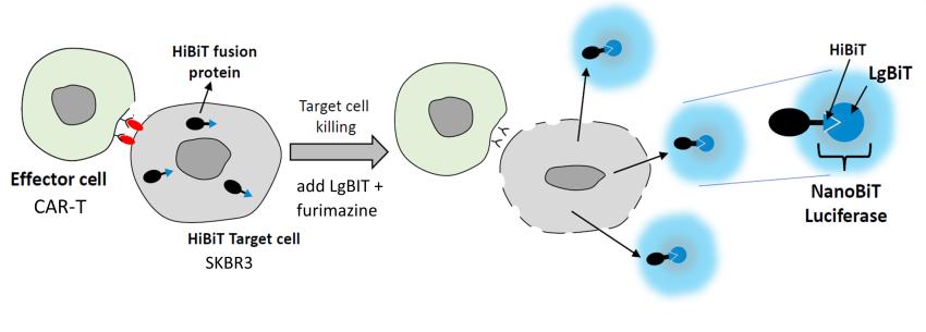
Figure 2. Promega HiBiT Techology (cell killing assay)
The HiBiT cell killing assay is based on a modified target cell line carrying the receptor of interest and the HiBit fusion protein. In presence of the effector CAR-T-cells the target cells become leaky and the furimazine substrate gets cleaved if HiBiT and LgBit form the active enzyme. The readout is luminescence.
All methods require thorough establishment to ensure that the suitable effector target ratio and incubation time is chosen for reproducible results. Due to the complex nature of CAR-T cells this can be tricky since the optimum conditions might vary from lot to lot, even with a well-controlled production process.
With that in mind reading the relevant guidelines obviously illustrate the hurdles that need to be overcome. The biological activity assay should have established and documented accuracy, sensitivity, specificity, and reproducibility and be amenable for validation. It should have pre-defined acceptance and rejection criteria. Therefore, it is important to plan for sufficient time for thorough assay development and understanding the capabilities and limitations of the method and the test item to set validation as well as assay acceptance criteria in a reasonable way. Another challenge is the requirement to include appropriate reference material and controls knowing that lots are limited in size.
Series logo designed by Chelsea Bates, Charles River Laboratories.