
Discovery
|
Julia Schueler, DVM
Macrophages as Double Agents
As immune cells macrophages fight infection. They also are a key driver in shaping the tumor microenvironment
The tumor microenvironment (TME) as an important player in regulating sensitivity and resistance towards cancer treatment has gained momentum in the last 15 years. But who knew that our own immune system was helping the TME do its job?
Aside from the stromal compartment–a market of the lethal tumor microenvironment—the immune cells infiltrating the tumor tissue are arguably the most important cell types shaping the characteristics of an individual tumor. This arsenal of immune cells, which should be protecting us against infection, instead engages in a steady crosstalk with the tumor cells.
The ability of the tumor to grow uncontrollably in a human body without being detected by the host immune system was first described as immuno-evasion by Douglas Hannahan, a scientist with the Swiss Institute for Experimental Research in 2011. Two major mechanisms lay below this phenomenon however: the absence of immune effector cells including cytotoxic T cells, and the presence of actively immune suppressive cells like regulatory T cells (Tregs) and myeloid-derived suppressor cells (MDSCs). Both can suppress cytotoxic lymphocyte´s activity.
How does the tumor microenvironment exploit macrophages?
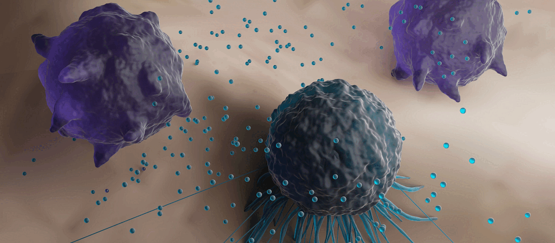
But none of these immune cells are quite as useful to the TME as a macrophage. These specialized immune systems engulf foreign substances harmful to us—which is they are often described as the garbage collectors of the immune system. They are capable of penetrating tissue and can reach out to any material our immune systems recognize as non-self.
But in conjunction with the TME, they, ironically, turn lethal. There are two subtypes of macrophages—M1 and M2—which differ in their physiological as well as their disease-related roles. The two subtypes are not strictly defined subtypes but rather fluidically transition between the two states, commonly called polarization.
The differentiation between M1 and M2 is valuable as it is basic and easy to apply: M1 activity inhibits cell proliferation and causes tissue damage while M2 activity promotes cell proliferation and tissue repair. This concept is similarly true for tumor progression; M1 macrophages are commonly considered as anti-tumorigenic, while M2 macrophages support tumor growth by contributing to many pro-tumorigenic processes like immune suppression, angiogenesis, tumor cell proliferation, metastasis and oxygen reduction. The TME as well as the tumor cells itself not only impact the polarization of already present macrophages, but they as also recruit them from the surrounding tissue.
So, understanding the principles and key players of TME-induced macrophage polarization is mandatory to understanding the pro- and anti-tumoral implications of tumor-associated macrophages (TAMs).
How can we model macrophages in the tumor microenvironment?
A detailed knowledge about the tumor-macrophage interaction helps us to discover innovative therapeutic approaches that manipulate this process. Mouse models are an excellent choice to test this interaction, as they include a fully functional immune system, including macrophages and other immune and non-immune cells present in the TME. Yet they still come with a number of drawbacks. Murine tumor models represent just a handful of tumor types and require surrogate markers. Plus, introducing human macrophages into an immune-compromised mouse is challenging as the currently available mouse strains do not support the engraftment of macrophages or enable the proper environment for their adaption into M1 or M2.
Some labs, such as ours, are using In vitro 3D models to study how macrophages interact with tumor cells in the TME. In vitro models offer a number of advantages:
- They are fully human as each cell type can be sourced from human donors and
- They offer the possibility of identifying critical cellular and molecular contributors to the disease by permitting manipulation of each in isolation.
In the context of macrophage-tumor interaction there are some factors that drive the translational value of such a 3D model: the triad of cell source, tissue architecture and technology. All build the basis for the different applications in drug discovery and tumor biology. By choosing patient-derived donor tissue and including stromal cells that shape the macrophage differentiation the biological relevance of the 3D model significantly increases Because these platforms mimic the TME very closely and can be used from target ID through preclinical efficacy to target immunosuppressive macrophages in tumors.
The ultimate proof of the translational value of these platforms is still missing. Not enough time or data is available to deliver fully approved drugs. Nevertheless, the that is available looks encouraging and the tremendous number of studies currently occurring now will enhance understanding the translational value of 3D models and the role of macrophages in tumor progression.
Attending AACR 2023? Check out our list of presentations and posters at this year's event.