
Discovery
|
Regina Kelder
Revolutionizing Oncology Drug Development
From implantable microdevices to 3D models, how two breakthrough tools make cancer treatments more effective—and meet the 3Rs objectives
This is part of Eureka's ongoing series, 4Rs: Looking Ahead Responsibly, which focuses on innovative projects, partnerships and collaborations that are helping to responsibly reduce, refine and replace research animals.
 There is no doubt that the arsenal of cancer drugs—from targeted therapies to immunotherapies to the up-and-coming gene and cell therapies—has never looked more promising. So why aren’t patient outcomes more favorable?
Immunotherapy is now the standard of care for several cancers, yet long-term survival with immunotherapy, while certainly evident in some advanced disease patients, is not a given. By one estimate, only about 15-20 percent of patients go on to achieve durable results. And while targeted therapies, from the 1997 gamechanger Herceptin to the first-ever KRAS inhibitor in 2021, can precisely kill the genes and proteins that help cancers grow and survive, many of these cancers are savvy enough to eventually develop resistance to these drugs.
Worse, most oncology drugs never even make it to market. By one estimate, the US Food and Drug Administration has reported a 97% failure rate at clinical trials for oncology drugs mostly due to drug efficacy (50%) and toxicity (30%) issues. And findings reported this year in JAMA estimated that failed drug development in oncology costs, at an industry level, an estimated $50 billion to $60 billion annually.
The reasons for failure are many, from drugs that don’t meet their targets to a need for better biomarkers and more translatable models. And while there is a plethora of well-established in vitro and in vivo models, they don’t always predict on their own what will happen in a patient.
Two companies, in very different ways, are trying to improve these dismal odds for cancer patients. Full disclosure, they also are strategic partners of Charles River Laboratories.
“With the discovery and development of novel agents that are intended to be used in combination either with other drugs or with other modalities, there is an increasing need for preclinical tumor models that can recapitulate the drug doses and schedules that will be used in patients,” said Julia Schueler, PhD, Research Director with Charles River’s Discovery Oncology business.
3D Models and Implantable Microdevices
The ideal model would contain the range of cell types typically found in human tumors including fibroblasts, endothelial cells and immune cells. The model would allow dosing of drug alone, in combination or in sequence in a manner resembling some of the dosing regimens used to treat cancer patients including continuous dosing schedules. Some of the models would be suitable for combinations with other modalities such as radiotherapy, as they represent tumor types that are usually treated with radiotherapy-containing regimens such as squamous carcinoma of the head and neck or rectal tumors.
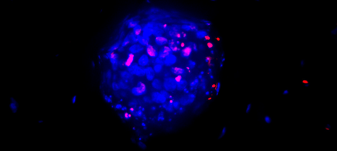
Cypre, a biotech company based in San Francisco, is breaking new ground in the development of 3D cancer models (shown in image above) that recapitulate the complex tissue in and around a tumor. Zeroing in on this ecosystem, or tumor microenvironment, allows researchers to gain new insights into a tumor’s playbook and to identify new therapeutic targets that might furnish a new generation of cancer drugs.
Kibur Medical, a Boston-based company, has developed implantable microdevice technology called the NanoNailTM that is about the size of a grain of rice and allows drug developers to design better tolerated, more potent oncology drugs or drug combinations and biomarkers predictive of drug efficacy, leading to an increased chance of clinical success.
While the goal of both Cypre and Kibur is to improve the oncology drug pipeline and ultimately help more patients, their technologies are also helping drug developers refine and reduce the use of animals in certain studies, a plus for 3Rs (the reduction, replacement and refinement of research animals.) Kibur’s implantable microdevices, which can rapidly screen the in vivo tumor molecular response to up to 20 drugs at one time, separately or in combination, in solid tumors require substantially fewer animals than conventional in vivo studies, said Colin Brenan, Chief Executive Officer of Kibur Medical.
Cancer technologies also focus on 3Rs
His company recently quantified this 3Rs impact in a clear cell renal carcinoma (CCRC) study conducted in partnership with the Brigham and Women’s Hospital in Boston and Charles River Laboratories’ Freiburg site, which specializes in PDX or patient-derived xenograft models.
The study exploited the power of the NanoNail for multi-factorial experiments to test a diversity of different single drugs and drug combinations in combination with a systemically delivered anti-PD1 treatment to identify potent combinations to which the CCRC responded since combination therapies have shown significant advances in treating later-stage or metastatic CCRC. A PDX from a 68-year-old man with clear cell renal carcinoma was implanted in 30 immunodeficient mice and Kibur then used the NanoNail to deeply profile the spatial phenotype and transcriptome in each tumor to 11 drugs administered simultaneously with/without the systemic anti-PD1.
Brenan said not only novel and unanticipated drug combinations were identified from this study, but the results were achieved with a six-fold decrease in the number of mice compared with the conventional approach for such a study. The reductions in animal use are potentially even greater for high-throughput, high-content screens that drug developers use early on for critical go/no decisions. “In these efficacy screenings you can measure maybe 60 drugs at a time,” said Brenan. “So, you are looking at a 20-fold decrease in the number of animals used.”
Cypre also impacts the use of animals, though in a different way. Its 3D in vitro platform creates patterned hydrogels in 96-well plates that encapsulate various cell types, from tumor and stromal cells to immune cells, in a scalable and automation fashion, says the company’s founder and CEO Kolin Hribar, PhD.
“What we have been able to see, and I think what differentiates our platform specifically, is the ability to more finely understand how novel therapeutics interact with the various tumor compartments, such as immune cells migrating through the stromal periphery of a tumor, the production of key cytokines, immune phenotype changes, and ultimately the reduction in tumor growth,” he said.
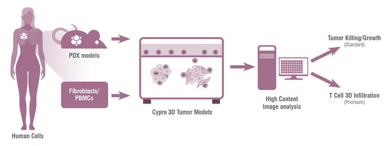
The technology also allows us to screen these effects at high throughput, said Hribar. “Cypre’s proprietary automation and analysis workflows help us build robust and standardized data packages across the genetic diversity of cancer subtypes, a digital tumor atlas if you will.” Ultimately, the company plans to use these datasets to train in silico systems to better interpret therapeutic efficacy and generate novel insights for biomarker and target discovery.
Which brings us to the 3Rs benefit.
Cypre’s 60+ solid cancer patient-derived 3D tumor models – 60 exclusively with Charles River and additional models in development —allow companies today to assess their compounds early on and determine which ones to move forward. There is also a small subset of PDX models for blood cancers. This early in vitro work can circumvent animal testing and strengthen the pathway from the lab to humans.
“We have preconfigured panels of these various models that are screened on a monthly basis,” says Hribar. “[A company] can just drop in their compound and … get data back in 30 days on the antitumor effects of their treatment.”
While the 3D models are currently used to refine or reduce use of animals, Hribar believes they could one day replace animals. “So, in one manner, we can use these systems [now] for disease modeling, and particularly for us, that is tumor modeling and understanding human tumor biology. But in the future, we could have a complete replacement of an animal … that functionally creates a representation of a human system with all the organs and integration of fluid flow.”
Toward regulatory and industry acceptance
Of course, to move forward drug developers must be willing to use the alternative technologies and incorporate the data in their regulatory filings. At the same time, regulators need to be open and receptive to include data generated with alternative models. By many indications change is happening in both camps.
In 2016, the US government included implantable microdose technology among the top ten list of the “most compelling research opportunities” that should be supported by the “cancer moonshot,” which was launched to accelerate scientific discovery, foster greater collaboration, and improve the sharing of cancer data. More recently, the 2022 FDA Modernization Act: 2.0 passed by Congress and signed by President Biden encourages the development of alternative methods by allowing researchers the opportunity to use the most rigorous scientific methods available, animal or not.
Brenan said currently their implantable technology is used for animal studies, and has received FDA approval for use in humans, but “the challenge is to clinically deploy the NanoNail to provide human tumor response data prior to an IND-filing. Having this information early in the development process could be potentially transformative by increasing success rates in the clinic.”
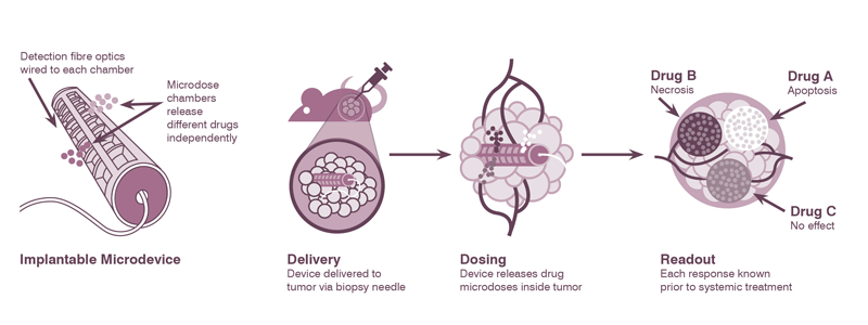
A study published earlier this year in Science Translational Medicine described using Kibur’s implantable device successfully in six patients undergoing brain surgery to remove a glioma, one of the few cancers whose prognosis has not improved over the years. The study provided clinical evidence that the drug-releasing microdevices can be used to safely and effectively obtain patient-specific, high-throughput biological data. The data can be integrated with, and is potentially superior to, other currently used biomarkers, the study said. The implantable device was used to test seven off-label drugs, from standard chemotherapies to targeted therapies, with some generating responses in patients.
“The study was small, but nonetheless super-encouraging, I thought, because it means there are alternative drugs out there that we haven’t previously considered that could be used to treat this devastating disease,” says Brenan. “It takes a long time to get these technologies adopted, but I would argue that change has to happen because we can't sustain this huge rate of clinical attrition from the current oncology drug development model.”
Hribar says 3D models, like Cypre’s, are the “here and now” for small molecules, antibodies, and cell therapies. In terms of reducing and replacing animals, Cypre has already started to see the effects of their data, he says.
“We have had clients use our system specifically to generate data that they can advance to IND, and in other cases, use our preconfigured Tumor Panels to refine their model selection for subsequent in vivo studies,” said Hribar. “In fact, I was surprised how quickly groups started to come to us in the last 8-9 months following passage of the FDA Modernization Act: 2.0, and say ‘Now that this Act is signed, can I just use your data for my pharmacology IND package instead of in vivo models’. We want to be cautiously optimistic on that concept because large regulatory changes take time, and we want to work closely with relevant agencies and our partners to ensure a smooth adoption for our clients.” Certainly, it seems that some advanced modalities like cell therapy favor this approach sooner than others, Hribar added.
In addition, Hribar said they will continue their march toward correlating responses between patient outcomes and their 3D model. “That's really the holy grail for validation work,” he said. The company published some of its work with Cedars Sinai Medical Center in 2019, showcasing the correlation of chemotherapy response in Cypre’s 3D in vitro models to the clinical outcome in glioblastoma patients, and more recently performed a correlative analysis with Charles River to compare 3D in vitro and in vivo PDX outcomes. “The correlation was clear for the subset of tested therapies and target classes. In other comparative tests particularly in the immuno-oncology setting, to my surprise, the 3D model showed a more human relevant TME by retaining key myeloid and human fibroblast cell types that were not necessarily preserved in the in vivo animal setting.”
That is indeed an exciting finding, and these early insights are prompting the company to expand its model database and pharmacology validation across indications such as non-small cell lung cancer, colorectal, renal, ovarian, gastric, and pancreatic to name a few.
From Schueler’s perspective, the growth of innovative tools and technologies is highly encouraging but adds that the field needs to be mindful about how they use them.
“During the last 15 years, a plethora of innovative preclinical drug testing platforms has been developed that offer the possibility of identifying critical cellular and molecular contributors to the disease,” says Schueler. “The major challenge now is to identify which assay and read-out combination, or combinations suits best the current scientific question or questions. A deep understanding of the different platforms along with their pros and cons is a prerequisite to make this decision.”