
Safety Assessment
|
Aleksandra Zuraw, DVM, PhD
Welcome to My World of Digital Pathology
Digital pathology can be an invaluable GLP-compliant system for learning and working fast, with real-time feedback
This is part of Eureka's ongoing series, Accelerating Innovation, about new technologies that are impacting the pace of drug discovery and development. Today's story is about digital pathology.
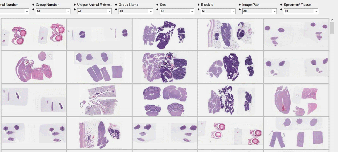
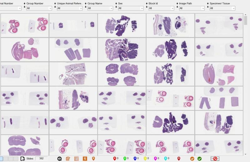
 I am sitting at a (relatively) clean desk, looking at my monitor and zooming in and out on high resolution digital images – whole slide images (WSI). For this study my microscope is resting under a dust cover. My glass slides were scanned with a whole slide scanner, and I am doing my evaluation on digital slides. This is what’s written in the study protocol, and I am happy not to have mountains of slide boxes surrounding me on my desk and around it. My workspace is limited to my computer and my monitor, and I love it!
Welcome to the world of full digital pathology where digital histopathology slide evaluation of entire studies is allowing us to be better pathologists. It’s so much easier to focus. No need to unpack any boxes, sort slides manually, mark them with Post It notes or with permanent markers for later additional review – I can do all this with a mouse click on my computer screen.
I can make a screenshot of an area on the slide I am uncertain about and send it to one of my more experienced colleagues in a split second via instant messaging. And if we need to discuss in more detail, I simply call them and share my screen or give them access to the study for their own digital review.
When I’m done for the day, I close the laptop and can actually use my home office to do some art activities with my kids – a nice change. Gone are the days where I have to lock my home office because of the boxes of slides on the floor during evaluation time.
What a positive difference to be able to evaluate slides digitally!
The work environment is more comfortable and the increase in the quality of work notable. The instant access to online consultations and the ability to leverage the collective pathology experience of our international pathology team (together we have over 150 pathologists with over 1,800 cumulative years of experience) is unmatched. This allows me to do my job in the most efficient and consistent way.
Taking digital pathology to a whole new level
Even though this new way of working is new for pathologists, digital pathology has been used by Charles River Laboratories for over a decade though recently we have taken it to a whole new level. We have achieved something no one in the drug development industry has done before – we validated our digital pathology systems for Good Laboratory Practice (GLP)-compliant primary histopathological evaluation.
This enormous cross-company team effort enabling our pathologists to work with digital slides, has allowed them to be more connected and supportive of each other that ever before in their quest to achieve the highest levels of quality for customers.
Knowing the actual and potential benefits of a new technology such as digital pathology often makes us overly enthusiastic. But in a GLP-compliant environment the enthusiasm and the willingness to adopt it is not enough. Every new method, every new computerized system (including a digital pathology system) needs to undergo a thorough multistep validation process that includes an IT, a regulatory and a scientific component.
During the digital pathology validation, the scientific component fell on the shoulders of the pathologists. We needed to make sure that everything that we would normally see on a glass slide was also visible on the whole slide image. We needed to know that for our intended use of diagnostic review the quality of the images produced by the whole slide scanner was equivalent to the quality of the images we were used to looking at with the microscope eyepieces.
To prove that we designed a special set of slides that covered everything we could potentially look at during a regular toxicology safety study evaluation.
We included slides from different species of laboratory animals, of different organs, different stains (including special chemical stains as well as immunohistochemistry markers), and slides with specific pathological changes .
From our previous work, we knew that digital pathology was a reliable method of slide evaluation. But including a set of slides with those specific, challenging lesions (a.k.a. critical features) mentioned in the literature and in a survey of over 150 toxicologic pathologists working at Charles River, and positively passing our computer systems validation tests gave us the necessary documentation required by GLP and the extra confidence of our pathologists and clients to deploy this solution across the company. We are sure digital pathology is good enough. We checked it!
But how rapid is digital pathology?
Ok, I hear your next question: We knew all along it was good enough, but is it fast enough? Does it match the speed of a microscope?
Everyone who has been doing digital pathology for several years, knows that this technology used to be painfully slow. Opening one image would take anywhere from 30 seconds to several minutes. This speed could not be further from the speeds at which an efficient, experienced pathologist would perform their toxicopathologic evaluation with their microscope.
In a fast-paced contract research organization (CRO) environment anything slower than a microscope is a no-go. The timelines of our sponsors depend on the speed of our evaluation. The evaluation takes as much time as necessary to achieve the highest quality, but not a second longer. Our digital pathology system had to meet this requirement and it did!
The technology enabling this unheard-of fast read is a combination of virtual cloud uploads and local laptop downloads. The study slides are uploaded to the cloud where they reside for the duration of the study and can be shared with the sponsor e.g., for peer review. During the actual evaluation pathologists download the WSI to their local computers. The computers are equipped with enough internal memory to store slides from several studies at the same time. Depending on the study size, the download can still take up to several hours, but it can be done overnight without the pathologist’s involvement and the slides are ready to be evaluated the next morning.
Digital pathology is an invaluable tool for working efficiently, flexibly, and collaboratively with a focus on our customer’s need for speed and quality.
Orchestrating the uploads and downloads with all the other study deadlines we are subjected to is not a trivial task, but we worked hard on it, and it is now an integral part of our workflow – the part of the workflow that helps us deliver the best quality of evaluation possible and fuels our capability to support around 85% of the FDA drug approvals happening every year.
Is it always seamless? No. But neither are other parts of our workflow. Nevertheless, we work hard together to make it happen and with every study it becomes easier and faster. We are working on automation that uses a combination of slide barcoding, AI-driven slide quality control, and software that can talk to each other, so we avoid manual tasks.
As any new technology digital pathology comes at a certain price and the question arises: Is this investment justified? How much better is it than what we were doing without it? Is the difference worth the price? This question comes up constantly but is not specific to digital pathology. It arises with implementation of any new technology.
I can speak to it from an early career pathologist. To me this investment is worth double, triple or quadruple the price. The speed of feedback I get from my colleagues when using digital pathology and instant communication media is unmatched even if we work in entirely different corners of the globe. My digital read gives the senior colleagues and peer reviewers the possibility to mark exactly (with digital pins and annotation on the WSI) the areas that need to be looked at more closely.
This is an invaluable tool for teaching, learning, and working fast with real time feedback. It takes the quality of our work to a whole new level and its value reaches far beyond the monetary investment necessary to put the infrastructure in place.
The slide digitization also opens the door to other methods that can help us support drug development even better. By converting the glass slides into digital images, we are generating a digital data point that can be integrated into an advanced digital data analytics pipeline.
We are now able leverage artificial intelligence and machine learning methods not only to perform image analysis based quantitative pathology, but also to build qualitative decision support tools for pathologists and build a foundation of multimodal predictive toxicology.
Does this seem farfetched to you? Maybe like science fiction? Let’s park this advanced vision for a second then…
If the only thing digital pathology gave you was the instant access to the cumulative pathology experience in your institution, would it be worth it? Add a tidy desk as bonus and I think you will know your answer. I know mine.
If you want more information about digital pathology, you will find our digital pathology educational resources here.