

Bridging the Gap Between Pathology and Computer Science
In this introductory video, Aleks explains how the idea for this educational interview series was born and her mission in doing so.

The Beginnings of Computational Pathology with Jeroen van der Laak
Special thanks to Radboud University Medical Center and Jeroen van der Laak, one of the leaders of the computational pathology group at Radboud UMC. In this first lecture, Jeroen and Aleks discuss:
- How the collaboration between computer scientists and pathologists started before computational pathology existed
- What defines success in the computational pathology field
- What changed in this field in the past 30 years
- Why being open-minded is one of the most important qualities in the digital pathology world

All About Whole Slide Images with Leslie Tessier and Daan Geijs
Aleks interviews anatomic pathologist Leslie Tessier and computer scientist Daan Geijs. Topics include:
- How is a glass slide created?
- How is it scanned and processed to become a whole slide image and to be viewed on a computer screen?
- What do pathologists and computer scientists need to know about those digital files to be able to work with them efficiently?
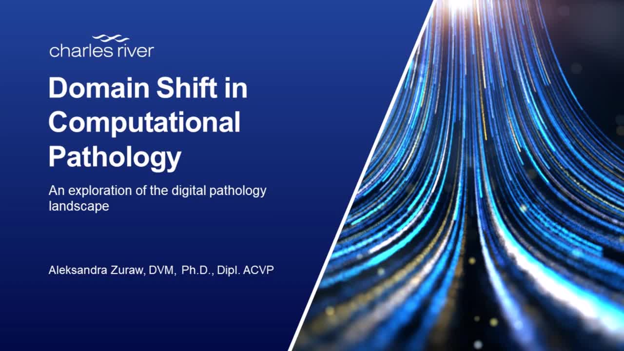
How to Deal with Domain Shift in Computational Pathology with Khrystyna Faryna
How do you build a robust computational pathology model if you have slides coming from all over the world, several different scanners, and even different times? The problem that needs to be tackled is domain shift. In this talk, Khrystyna Faryna, a PhD candidate at the Radboud UMC computational pathology group, will answer your questions related to domain shift:
- What is a domain in the context of computational pathology?
- What kind of domains do we have?
- Why is it important to account for domain shift?
- Can we make a model “domain shift proof?”
- What are the methods used to make a model robust to domain shift?
- How robust can it be?

Model Performance Metrics with Francesco Ciompi and Leander van Eekelen
When developing an image analysis model it is crucial to benchmark it, to check if it is performing well. Benchmarking requires us to ask:
- How do you know if your model is performing well?
- What model performance metrics are relevant for different computer vision tasks?
- How do we divide our image data to not only develop, but also test and validate, our models?
- What is cross-validation? When should it be used and how?
- What does it mean to say that a model is working well?
Aleks asks Francesco Ciompi and Leander van Eekelen to explain. Listen to this webinar-style presentation and choose the right metrics to check the performance of your model.

Computer Vision Approaches Used in Tissue Image Analysis with Leander van Eekelen
Different pathology problems require different computer vision approaches to design optimal tissue image analysis models. But what are the correct approaches?
Aleks discusses the following questions with Leander van Eekelen from the Radboud UMC Computational Pathology Group:
- How is working with whole slide images similar to working with natural images, and how is it different?
- Classical image analysis versus AI and deep-learning based image analysis - what are the similarities and differences?
- What computer vision tasks are used for analyzing images?
- What is:
- Image classification
- Object detection
- Semantic segmentation
- Instance segmentation
- Panoptic segmentation

Deep Learning for Tissue Image Analysis with Meyke Hermsen
Deep Learning has been successfully used for analyzing pathology images. What exactly is it and what are the different types of deep learning used in tissue image analysis? How does supervised deep learning work and what are the current applications in pathology?
Meyke Hermsen, a computational pathology PhD candidate at Radboud UMC, will answer these and many other questions.

Weakly Supervised Deep Learning with Daan Geijs
In this talk with Aleks and Daan, you will learn:
- What is weakly supervised deep learning?
- What labels can be used for weakly supervised deep learning?
- What methods of weakly supervised deep learning are currently being used?
- MIL
- CLAM
- Streaming
- How does the performance of those methods differ?
- What are the current applications of weakly supervised tissue image analysis methods in pathology?

Unsupervised Deep Learning Tissue Image Analysis with Geert Litjens
Can fully unsupervised deep learning be used in pathology? Can we trust it? How mature is it?
In this episode Geert Litjens, a professor in the Computational Pathology Group at the Radboud UMC in Nijmegen, The Netherlands, will introduce you to the world of unsupervised deep learning for tissue image analysis.
Aleks and Geert discuss:
- What is unsupervised deep learning and how exactly does it differ from supervised deep learning?
- What are the advantages and disadvantages of unsupervised learning in tissue image analysis?
- What are the current methods used?
- What are the current applications of unsupervised deep learning to tissue image analysis?

Model Uncertainty in Computational Pathology
We all look up the weather forecast every day, although we know it is not guaranteed to be correct – we accept it, while often secretly hoping the predictions don't come true.
But how should we deal with computational pathology model uncertainty? How much uncertainty can we afford? How do we deal with different uncertainty levels, and when should we say "enough is enough" and disregard the prediction altogether?
In this talk, expert Milda Poceviciute from Linköping University will walk you through the nuances of model uncertainty. She will explain:
- What is uncertainty?
- Why is it important to analyze uncertainty?
- What is uncertainty analysis used for?
- Uncertainty versus attention vs probability of a model
- What are the applications of uncertainty analysis in pathology?

Intersection Between Histopathology and Spatially Resolved Gene Expression
Predictions of molecular properties of tissue from histopathology images is a very promising method to reduce cost and increase the accessibility of cancer patient care. A lot has been published on the subject and the results have been surprisingly good, but how can we verify which image features actually correspond to the molecular properties?
We can do this through Spatially Resolved Gene Expression.
Aleks and her guest, Dr. Eduard Chelebian, discuss:
- What is spatially resolved gene expression?
- Why is it important to relate histopathology and gene expression?
- How is it done?
- What are the current applications for this technology?

How to Make AI Outputs Convincing for Users in Assisted-Reading Setups
In the final video of this series, Aleks and guest Leslie Tessier discuss how to make AI outputs convincing for users in assisted-reading setups and increase its adoption.
Listen to perspectives of a veterinary surgeon and medical doctor specialized in pathology.
About the Presenter
Aleksandra Zuraw, DVM, Ph.D., Dipl. ACVP
Veterinary Pathologist II
Charles River
A veterinary pathologist by training, Aleksandra Zuraw is particularly passionate about digital pathology. Based at Charles River’s Pathology Associates site in Frederick, MD, she has extensive experience in digital pathology and image analysis. Explore her blog.
Webinar Series: Are You Ready for the Digital Pathology Revolution?
Digitalization is revolutionizing traditional laboratory pathology. Potentially more objective, accurate, and faster than traditional microscopy, this technology is changing our workflow.
Watch now