
Assess Mode of Action and Dose Toxicity with Cell Painting in Drug Discovery
Cell painting in drug discovery is a novel unbiased method to investigate subtle changes in cellular homeostasis, which rapidly produces qualitative data demonstrating the effect of a compound and/or dose toxicity on a target at the single-cell level. The strategy involves immunohistochemistry application of six dyes across five channels, revealing eight major cellular organelles. The image-based profiles can be acquired by extracting >3000 morphological features at the single-cell level and identify subtle changes in cellular morphology in response to treatments.
Compatible with a board range of modalities, cell painting allows for unbiased insight into potential toxicity and dose toxicity. Cell painting data provides a mechanistic understanding of your compound's mode of action within a single cell, and the end-point biological effect of your compound. This investigative method can help filter and refine large volumes of candidate data generated during the drug discovery process, and pairs well with a broad range of studies. Following lead optimization, cell painting for the drug discovery process can be leveraged to build an understanding of how your compound reacts and functions.
Our cell painting assay service is fully compatible with high-throughput read-out and analysis, and is based on the pipeline developed by JUMP-Cell Painting Consortium.
How Cell Painting Assays Supports Drug Development Research
Cell painting assays can be used in drug development to evaluate the effects of compounds on cellular morphology and to identify potential drug candidates.
Phenotypic Screening
Cell painting assays can be used to screen large compound libraries to identify those that induce specific changes in cellular morphology associated with disease. The assay can cluster hits into groups with biologically similar effects. Based on the similarity to the reference compounds it can reveal a compound’s mechanism of action (MOA) and simultaneously uncover previously unsuspected off-target activities.
The method is also applicable in rare disease e.g., n=1 repurposing studies, where cell painting could identify compounds able to rescue the disease specific phenotypes.
MOA Studies
Once a compound has been identified as a potential drug candidate, cell painting assays can be used to determine its mechanism of action. By comparing the changes in cellular morphology induced by the compound to a database of known MOA, researchers can infer how the compound is modulating cell biology.
Toxicity Testing
Cell painting can also be used to evaluate the toxicity of compounds by comparing the changes in cellular morphology induced by a compound to a database of known toxic effects.
Combination Therapies
Cell painting assays can be used to identify synergistic combinations of drugs that induce specific changes in cellular morphology associated with disease. This approach can identify new drug combinations that may be more effective than single drugs alone.

Novel In Vitro Strategies to De-Risk Small Molecule Candidate Selection
In this webinar, explore how novel technologies are elevating de-risking during drug discovery, and how and when to apply these approaches to your pipeline.
Watch Now
Cell Painting Methodology
Cell painting, first developed by the Broad Institute of MIT and Harvard, can be added to your pipeline as a standalone study or as an integrated study within a range high content screening services. Sourced human cells are stained with six dyes and assessed using the standardized quantification workflow as published by the JUMP-Cell Painting Consortium. The cell painting method involves a series of steps, including:
1. Cell preparation: Cells are seeded onto a plate or slide and allowed to grow to a desired density.
2. Fixation: Cells are fixed with a chemical fixative such as paraformaldehyde to preserve their morphology.
3. Staining: Cells are stained with a set of fluorescent dyes that bind to specific cellular components, such as the nucleus, cytoskeleton, and mitochondria. The dyes used in cell painting are selected to provide a comprehensive view of cellular morphology and organization.
4. Multiplex imaging services: Stained cells are imaged using automated microscopy. Multiple images are typically acquired for each sample to capture the full complexity of cellular morphology and organization.
5. Multiplex image processing: The resulting multiplex images are processed using software to segment the cells and extract features that describe various aspects of their morphology, texture, and organization.
6. Data analysis: The extracted features are analyzed using machine learning algorithms to identify patterns and changes in cellular morphology that are associated with disease or drug treatment.
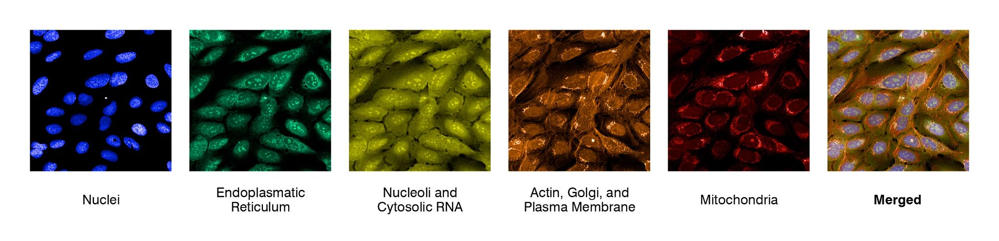
In this example, eight cellular organelles were visualized through five fluorescent dyes. Images were taken on a Yokogawa CellVoyager 8000 spinning-disk confocal platform using a 60x water immersion:
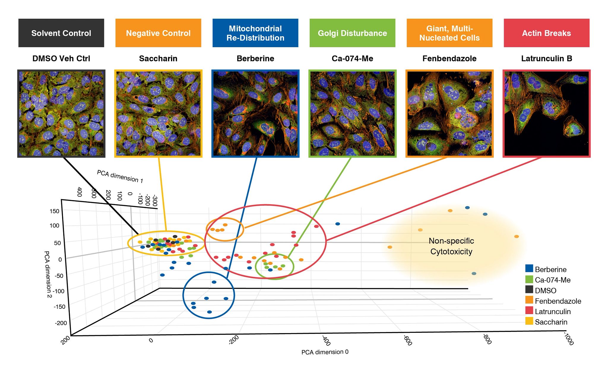
Reference chemicals shows distinct morphological phenotypes of compound treatment after 48hrs treatment (selected chemicals are displayed). (B) PCA plots confirm distinct phenotypic effects, but also show non-specific cytotoxicity at higher concentrations:
Assessment via cell painting assay provides an excellent and rapid route to understanding the mode of action of your compounds, and refining lead selection by removing candidates with potential toxic liability from your drug discovery development pipeline.
Evaluating the Cell Painting Assay in the Early Drug Discovery Pipeline to Reveal Mechanistic Properties of Test Compounds
Learn about the correlation between the dose-dependent effect of test compounds and the mechanism of action (MOA) predicted by a cell painting assay.
Download the Poster
Frequently Asked Questions (FAQs) About Cell Painting Assay
-
What type of cells can be used in cell painting?
The cell painting method for drug discovery can be applied to many different types of cells. The choice of cell type depends on the specific research question being addressed. For example, if the goal is to study the effects of drugs on cancer cells, cancer cell lines such as HeLa or MCF-7 may be used. If the goal is to study hepatotoxic effects of drug candidates, hepatocyte derived cell lines such HepaRG or HepG2 are more relevant.
It is important to note that different cell types may require different staining conditions and imaging parameters. Therefore, it is important to optimize the staining and imaging protocols for each cell type to obtain quality data. Additionally, it is important to consider the appropriate controls to ensure that any changes observed in cellular morphology are specific to the experimental conditions being tested.
-
What are the reference compounds in cell painting?
Reference compounds in the cell painting assay are a set of known compounds with well-defined MOA that are used to establish a baseline for comparison with experimental compounds. The reference compounds are typically selected to represent a diverse range of biological activities and pathways, and may include drugs that target the cytoskeleton, microtubules, mitochondria, and other cellular components.
The reference compounds used in the cell painting assay may vary depending on the specific research question. For example, if the goal is to identify drugs that induce cancer cell death, the reference compounds may include chemotherapeutic agents that are known to be effective against cancer cells. If the goal is to identify drugs that induce changes in mitochondrial morphology, the reference compounds may include drugs that specifically target mitochondrial function.
-
Can cell painting identify the mode of action of an unknown substance?
Yes, the cell painting assay can provide information about the mode of action of a molecule by identifying and analyzing changes in cellular morphology and organization that are associated with specific biological pathways and processes.
The cell painting assay data can also be used to predict the mode of action of molecules based on their similarity to known reference compounds with well-defined mechanisms of action. For example, using the right reference compounds such as Berberine, cell painting can distinguish between microtubule-disrupting agents and agents that affect other cellular structures based on their effects on cellular morphology and organization.
-
What is the Jump Cell Painting Consortium?
The Jump Cell Painting Consortium is a collaborative effort aimed at advancing the use of the cell painting assay in drug discovery and toxicology research. The consortium was launched in 2018 by the University of California, San Francisco (UCSF), the Chan Zuckerberg Biohub, and the Allen Institute for Cell Science, with funding from the JPB Foundation.
The goal of the consortium is to develop standardized protocols and data analysis methods for the cell painting assay, and to promote the use of the assay in large-scale drug discovery and toxicology screening. The consortium brings together academic and industry researchers, bioinformaticians, and technology providers to share data, methods, and expertise, and to collaborate on research projects.
The consortium has developed a standardized cell painting protocol and data analysis pipeline, and has generated a large-scale dataset of cell painting images and metadata for thousands of compounds across a variety of cell types and experimental conditions. The dataset can be used to train machine learning models and to test the performance of new analysis methods.
Jump Cell Painting Consortium represents a major effort to promote the use of the cell painting assay in drug discovery and toxicology research, and to accelerate the development of new treatments for human diseases.
-
What is cell painting?
The current form of cell painting method was developed by Anne Carpenter and her colleagues at the Broad Institute of MIT and Harvard in the early 2000s. The method involves staining cells with a set of fluorescent dyes that bind to specific cellular components, such as the nucleus, cytoskeleton, and mitochondria. These stained cells are then imaged using automated microscopy, and the resulting images are analyzed using machine learning algorithms to extract features that describe various aspects of the cells' morphology, texture, and organization. Cell painting is now widely used in drug discovery and other areas of cell biology research.