Nonclincal and Clinical ELISpot Analysis
ELISpot assays are highly sensitive immunoassays that measure the frequency of antigen-specific T cells by measuring the number of cytokine-secreting cells within the blood at a single cell level. B cell ELISpot assays can also be used to quantify the number of antigen-specific B cells that have recently been activated in vivo or to quantify memory B cells which have been activated with a specific stimulus ex vivo.
Advancing Studies from Discovery to the Clinic with ELISpot Assays
Accurate measurement of antigen-specific immune responses is essential for evaluating the safety and efficacy of new vaccines, gene therapies, and immuno-oncology treatments.
ELISpot assays address the challenge of detecting rare, cytokine-secreting T and B cells, providing sensitive, single-cell resolution data that other platforms may overlook. Sponsors who partner with us gain access to validated assays, regulatory expertise, and integrated biomarker services that translate complex immune data into actionable insights.
Solve the Challenge of Rare Cell Detection
ELISpot provides single-cell sensitivity to detect low-frequency immune responses, enabling critical insights into therapeutic impact early in development.

Translate Immune Monitoring into Development Decisions
We integrate ELISpot assays within a broader portfolio, helping you design, validate, and interpret immune monitoring strategies to accelerate decision making in programs.
Webinar: The Critical Role of Bioanalysis in AAV Therapies
This webinar will explore the expanding role of bioanalysis in AAV therapies, focusing on biodistribution, transgene expression, immunogenicity, and ELISpot.
Watch Now
Antigen-Specific T Cell ELISpot Assay Protocol
ELISpot analysis can be performed for a single cytokine or combinations of up to five cytokines by following the ELISpot assay protocol described below.
Cell Culture
For the cytokine(s) of interest, fresh or thawed peripheral blood mononuclear cells (PBMCs) or other relevant tissue cells are cultured on a membrane surface coated with a specific capture antibody in the presence or absence of antigenic stimuli.
Cytokine Capture
The cytokine(s) secreted by the cells after the stimulation is thus captured onto the membrane surface.
Cell Cycle and Proliferation
Determine cell cycle phases and monitor proliferation rates to evaluate compound effects on cellular growth and division.
Cell Removal and Cytokine Detection
After an appropriate incubation time, cells are removed, and the secreted cytokine(s) are detected using a detection antibody.
Binding via HRP, ALP, or Fluorescent Readout
The binding is captured using enzymatic (horseradish peroxidase [HRP] or alkaline phosphatase [ALP]) or fluorescent readout, where spots become visible.
Plate Washing
The plate is then washed to stop the reaction, and the membrane is allowed to dry before analysis.

eGuide: Biomarker Solutions from Discovery to Clinical Trials
Learn how strategic biomarker integration can accelerate research and de-risk pipelines to bring life-saving therapies to patients faster. This guide helps you uncover smarter ways to de-risk studies and accelerate timelines, without compromising scientific rigor.
Read the Guide
Why Use ELISpot Assays?
ELISpot assays provide a powerful readout of antigen-specific immune responses, making them a key tool in vaccine development, gene therapy, and immuno-oncology. By detecting cytokine secretion at the single-cell level, ELISpot delivers unmatched sensitivity to capture even low-frequency T cell and B cell responses.

Highly Sensitive Detection
Measure rare, antigen-specific cells with precision, enabling early insights into immune response dynamics and therapeutic impact.

Flexible Applications
Deployable across early-phase clinical trials, vaccine studies, and gene therapy programs, ELISpot assays adapt to a wide range of therapeutic modalities.

Clinically Relevant Readouts
Generate data that supports decision-making by tracking immune activation and guiding therapeutic optimization.

Expert Scientific Guidance
Our ELISpot experts work with you to design and interpret studies, ensuring assay strategies are aligned with your development goals.
ELISpot Assay Validation
Prior to ELISpot analysis of nonclinical and clinical samples, a fit-for-purpose validation is designed based on the logistics of each trial, along with the intended purpose of the data. For clinical trial analysis, blood is drawn from subjects and is then transported to either one of our laboratories or a local laboratory for PBMC isolation within an appropriate timeframe. PBMCs may be analyzed immediately or frozen and stored in liquid nitrogen until ELISpot testing.
Clinical Sample ELISpot Assay Protocol
Validation of key elements for pre-analytical sample preparation and ELISpot setup is critical prior to analysis of clinical samples for each cytokine or assay. This includes criteria like critical reagents, validating operators, and testing parameters, including intra-assay and inter-analyst/assay precision, linearity, stability, and LOD/cut-point calculations.

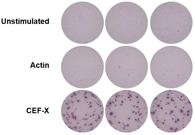
Representative Images from Healthy Donor PBMCs
Representative data is shown below from a validation study to assess IFN-γ secreting cells in response to the CEF-X, a positive control composed of a pool of viral peptides including CMV, EBV, and influenza virus.
PBMCs from healthy donors were isolated and frozen down to mimic clinical trial logistics. The healthy donor PBMCs were then established in culture alongside commercially available frozen quality control (QC) PBMCs and challenged with media (unstimulated), a negative control peptide pool (Actin), and a positive control peptide pool (CEF-X).
Intra- and Inter-Donor Variability Determined Through ELISpot Analysis
Some variation is also expected in the magnitude of the response between different donors. Once it has been deemed fit for the intended purpose, it can be applied to the investigation of samples from clinical trial subjects.

Frequently Asked Questions (FAQs) for ELISpot Assay Services
-
What are some ELISpot assay applications?
Key ELISpot assay applications include monitoring immune responses during preclinical testing and clinical trials, ex vivo stimulation of immune cells to measure frequency of cells specific to the antigen of interest, and vaccine efficacy.
-
What are the benefits of using ELISpot assays?
One of the main benefits of ELISpot assays is that they are extremely sensitive for the detection, measurement, and analysis of rare, antigen-specific T cells and B cells and because of its high sensitivity, it can detect and enumerate a single cell that secrets a protein of interest such as a cytokine. It also provides an indication of the type of cytokine response that has been elicited and hence the type of immune response.
-
What can influence ELISpot assay performance?
ELISpot assay performance can be influenced by several different factors, including cell recovery and viability, operator technique and proficiency, and result variability.