
Parkinson’s Disease Animal Model Studies
We offer multiple animal models of Parkinson’s disease to support your research and Parkinson's drug discovery. Our scientists use 6-hydroxydopamine (6-OHDA) and MPTP, the most popular neurotoxin animal models of Parkinson's disease. Since 10% of all Parkinson's disease cases are genetic in nature, we also employ the more common genetic Parkinson's disease mouse models. All our animal models of Parkinson’s disease track induction, behavioral deficits, and monoamine levels and can be further investigated with a wide range of behavior, imaging, and bioanalysis techniques outlined below.
Parkinson's Disease Research Studies
Our Parkinson’s disease animal models have been validated using multiple endpoints, which can be combined in your study to determine drug efficacy against Parkinson’s disease pathologies. These include locomotion function, kinematic gait analysis, in vivo imaging, and analysis of biomarkers such as α-synuclein, tyrosine hydroxylase, NfL, and markers of neuroinflammation. Our neuroscience experts can guide you to select the most appropriate model, readouts and endpoints, and study design to generate the actionable data you need to advance your program forward.
Features of Animal Models of Parkinson’s Disease
MPTP Mouse Model of Parkinson's Disease
The MPTP mouse model is a chemically induced animal model of Parkinson's disease and is one of the most widely used Parkinson's disease animal models. The model is characterized by decreased dopamine, DOPAC, and HVA content in the striatum, as well as loss of dopaminergic neurons in the substantia nigra.
- Fine motor and kinematic gait analysis shows changes in interlimb coordination, speed of movement, hind limb joint angles and limb trajectories
- PET imaging with dopamine transporter (DAT) ligands shows decreased DAT density, while imaging with 18F-FDG shows decreased glucose metabolism
- Immunohistochemistry confirms loss of TH-positive cells and increased neuroinflammation in the substantia nigra
-
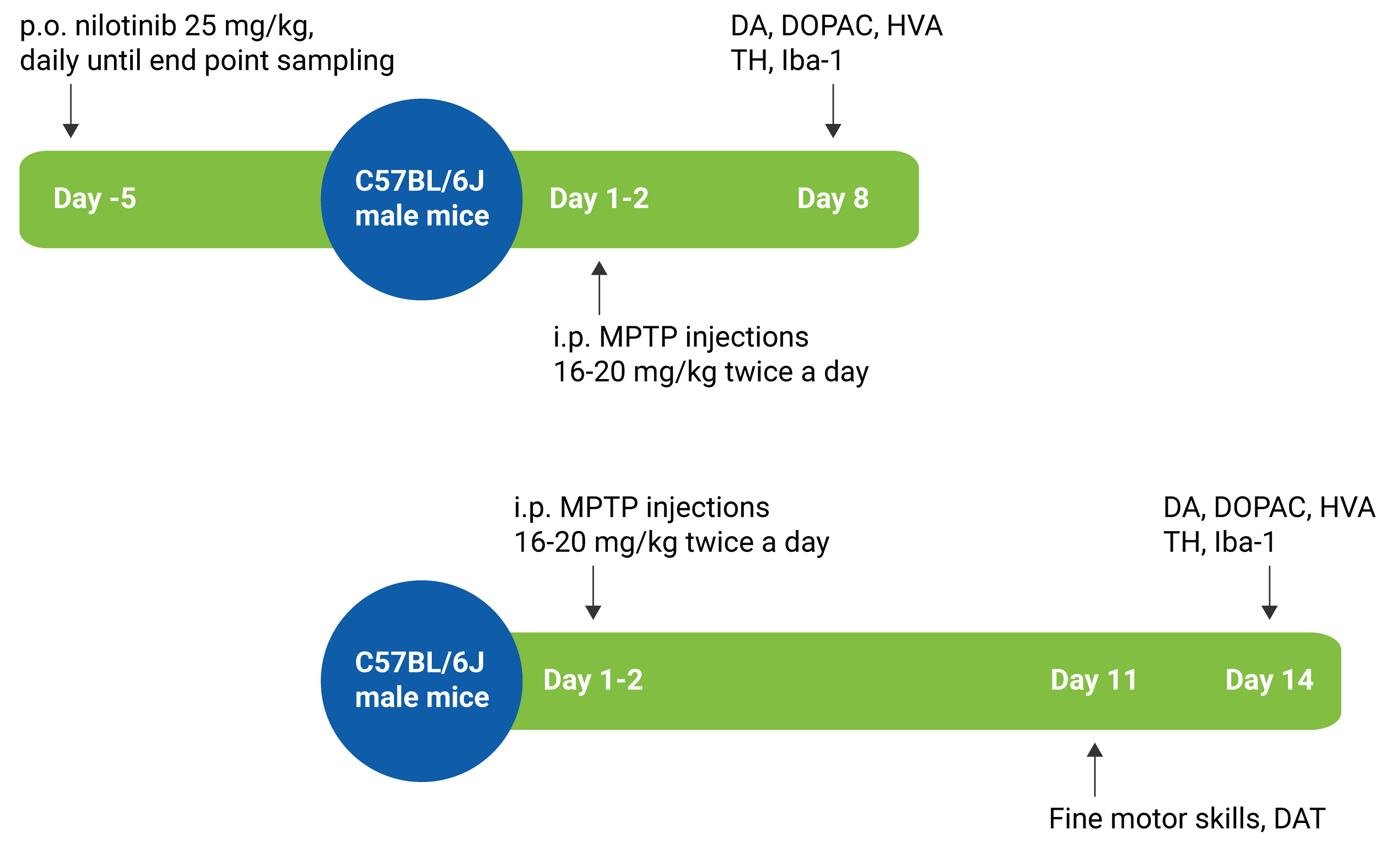
MPTP Mouse Model Study Paradigm
-
MPTP Mouse Model Validation Data
MPTP Mouse Model: Neurochemistry
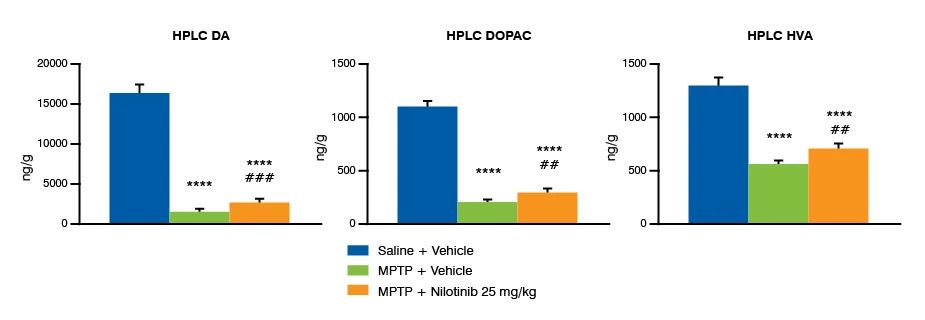
MPTP produces a robust reduction in levels of striatal dopamine (DA) and its metabolites DOPAC and HVA measured with HPLC. Treatment with the tyrosine kinase inhibitor nilotinib 25 mg/kg can partially rescue the MPTP-induced reduction of DA and DA metabolites. ***p<0.001, ****p<0.0001 MPTP - Vehicle vs. Saline - Vehicle. ##p<0.01, ###p<0.001 MPTP – Vehicle vs. MPTP - Nilotinib (Welch’s t-test). Data are presented as mean + SEM.
MPTP Mouse Model: Tyrosine Hydroxylase (TH) Staining In The Substantia Nigra Pars Compacta (SNpc)
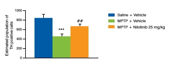
MPTP injections lead to a decrease in the number of TH-positive cells in the SNpc of mice. The tyrosine kinase inhibitor nilotinib can partially reverse the effect of MPTP. *** p < 0.001 MPTP + Vehicle vs. Saline + Vehicle. ## p < 0.001 MPTP + Vehicle vs. MPTP + Nilotinib. (Welch’s t-test). Data are presented as mean ± SEM, n=15/group.
6-OHDA Rat Model of Parkinson's Disease
The 6-OHDA rat model is a chemically induced Parkinson’s disease animal model that leads to loss of dopamine cells in one brain hemisphere. Our validation data show that the 6-OHDA model has decreased dopamine and dopamine metabolites (DOPAC and HVA) in the striatum, as well as loss of dopaminergic neurons. Motor deficits in this model can be investigated using fine motor analysis of gait and balance.
- Alterations in gait, including changes to forelimb movements, interlimb coordination and knee and ankle angles
- Detection and confirmation of lesion with MRI
- PET imaging with dopamine transporter ligands
- Decreased dopamine and metabolites in striatal tissues
-
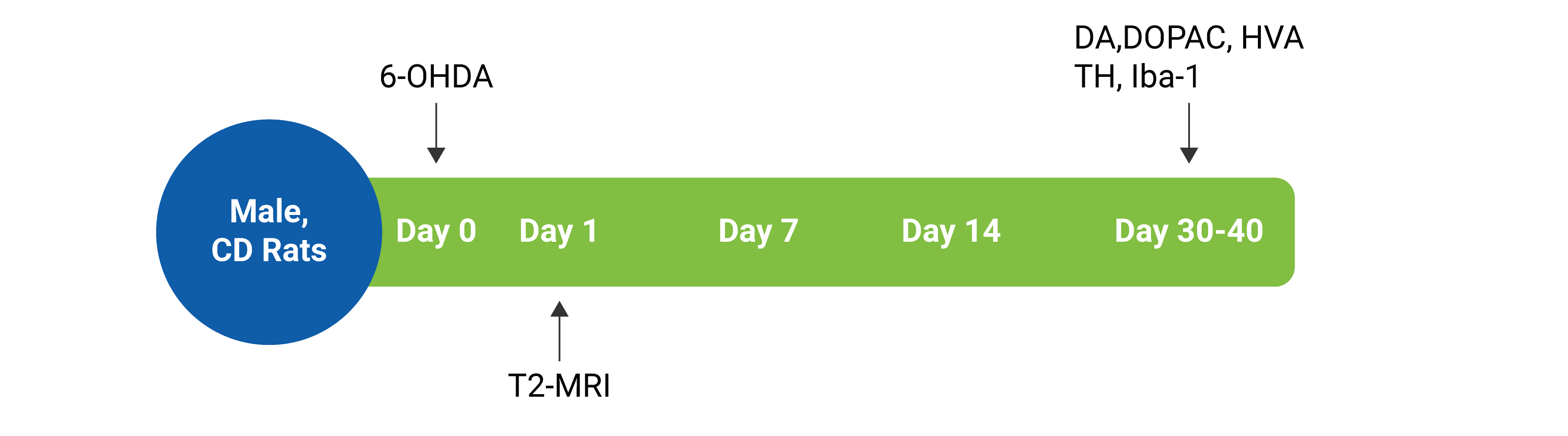
6-OHDA Model Study Paradigm
-
6-OHDA Model Validation Data
Monoamine Levels In The 6-OHDA Rat Model
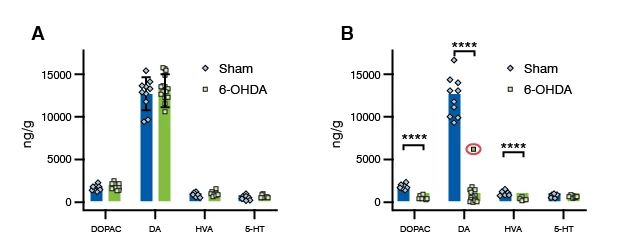
Total dopamine (DA), DA metabolites DOPAC and HVA, and serotonin (5-HT) levels in the striatum approx. 5 weeks after a unilateral injection of 6-OHDA. (A) Sham and 6-OHDA rats have similar monoamine levels in the control brain hemisphere, (B) In the injected hemisphere, 6-OHDA leads to a robust decrease in striatal concentrations of DA and DA metabolites. Incomplete lesion highlighted with a red circle .
6-OHDA Rat Model Immunohistochemistry: Tyrosine Hydroxylase (TH) and Staining in Substantia Nigra
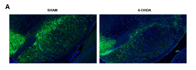
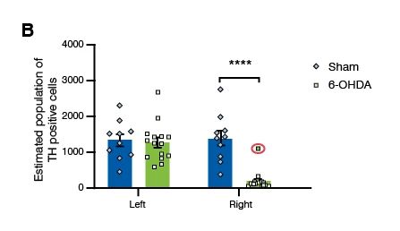
Figure A: A decrease in TH-positive cells in the substantia nigra pars compacta (SNpc) of 6-OHDA-injected animals can be visually observed in the sections. Figure B: Estimated TH-positive cell count in the SNpc approx. 5 weeks after lesioning show a marked loss of TH positive cells in the injected right hemisphere of rats that received 6-OHDA (****p<00001, multiple t-test, n = 10 SHAM, n = 15 6-OHDA. Incomplete lesion highlighted with red a circle.
6-OHDA Rat Model: In Vivo Imaging
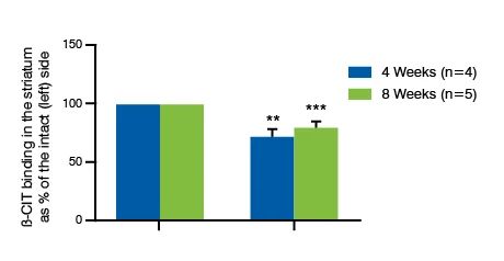
Injection of 6-OHDA leads to an approx. 20% decrease in β-CIT binding to dopamine transporters (DAT) on the lesioned side (right) as compared to the intact side (left) four and eight weeks post-lesioning (**p < 0.01, ***p < 0.001 as compared to left side, Welch’s t-test; data shown as mean ± SD
6-OHDA Rat Model Lesioning

Alpha-Synuclein Mouse Model of Parkinson's Disease
The AAV-A53T Alpha-Synuclein animal model is available in both rats (male CD rats) and mice (male C57BL/6J mice). It is a biological transduction model in which overexpression of A53T alpha-synuclein in the substantia nigra is induced by unilateral stereotaxic injection of AAV vectors. Our validation data show that this genetic Parkinson's disease animal model develops alpha synuclein pathology with reduction of striatal dopamine levels, loss of dopaminergic neurons, and neuroinflammation in the substantia nigra. Motor, gait, and balance analysis shows that fine motor skills are significantly affected in the alpha-synuclein mouse model.
Alpha-synuclein mouse model
- Significant deficits in motor, gait, and balance performance
- Loss of dopamine in the ipsilateral striatum
- Loss of TH-positive cells and increased neuroinflammatory markers in the ipsilateral striatum
Alpha-synuclein rat model
- Asymmetric gait and motor changes
- Unilateral reduction in dopamine and its metabolites in striatal tissue
-
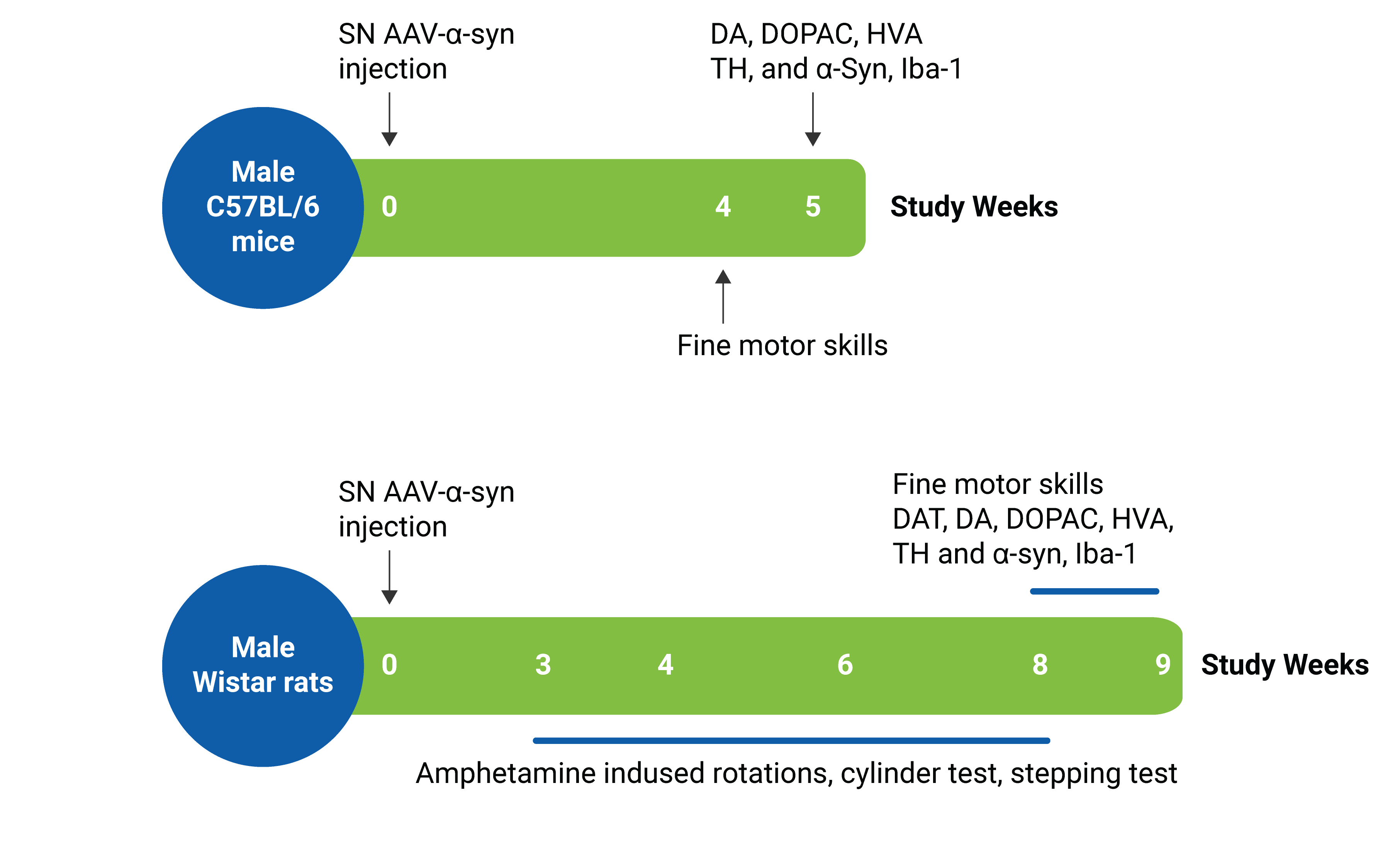
Alpha-synuclein Mouse Model Study Paradigm
-
Alpha-synuclein Mouse Model Validation Data
AAV-A53T Alpha-synuclein Mouse Model: Alpha-synuclein Immunoreactivity
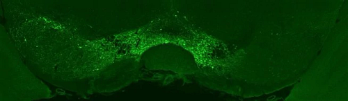
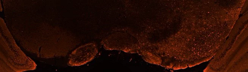
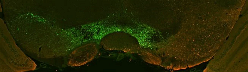
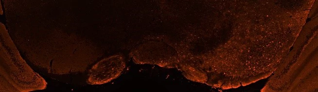
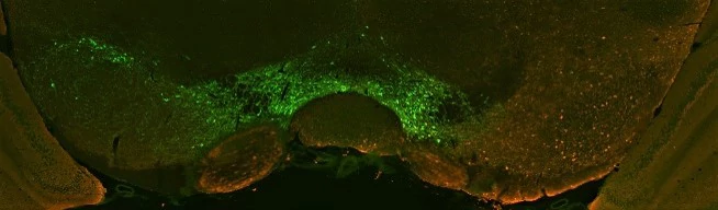
Immunofluorescence staining of tyrosine hydroxylase (TH, top image) and alpha-synuclein (middle image), and TH+ alpha-synuclein (bottom row) in the substantia nigra of rats injected with either AAV-A53T Alpha-Synuclein or a control AAV-Null vector. Alpha-synuclein immunoreactivity is observed in the AAV-A53T Alpha-Synuclein injected treatment group.
AAV-A53T Alpha-synuclein Mouse Model: Tyrosine Hydroxylase (TH) Staining in the Substantia Nigra Pars Compacta (SNpc)
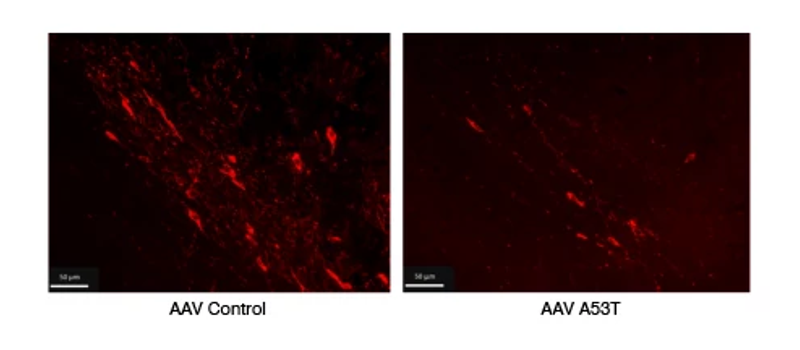
Figure A: An injection of AAV-A53T Alpha-Synuclein into the substantia nigra (SN) leads to a loss of TH-immunoreactive cells in the SNpc nine weeks post-injection (rats). Figure B: Analysis of number of TH-immunoreactive cells in the SNpc shows a decrease in cell number in the injected (ipsilateral) side. ** p < 0.01; *** p < 0.001, **** p < 0.0001. AAV control vs. AAV A53T (Welch’s t-test). Data are presented as mean + SEM./p>
AAV-A53T Alpha-synuclein Model: Neuroinflammation
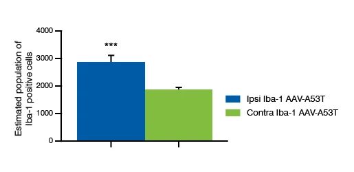
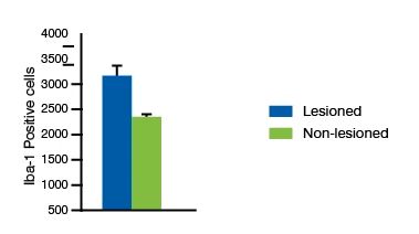
Immunostaining and quantification of Iba-1, a marker for microglia, shows an increase in Iba1-immunoreactive cells in the injected, ipsilateral SNpc five weeks after AAV-A53T Alpha-Synuclein delivery. *** p < 0.001, Ipsilateral vs. contralateral side (Welch’s t-test). Data are presented as mean + SEM, n=15.
PINK1/PARK2 Knockout Mouse Model
Mitophagy, the removal of damaged mitochondria, is a key pathological process in Parkinson’s disease and is being investigated as a therapeutic target. PINK1 and Parkin proteins, encoded by PINK1 and PARK2 genes respectively, have been linked to familial Parkinson’s and are vital to mitophagy processes. Our scientists have validated a PINK1/PARK2 double knockout mouse model, which can be used in efficacy studies for novel Parkinson’s disease therapies.
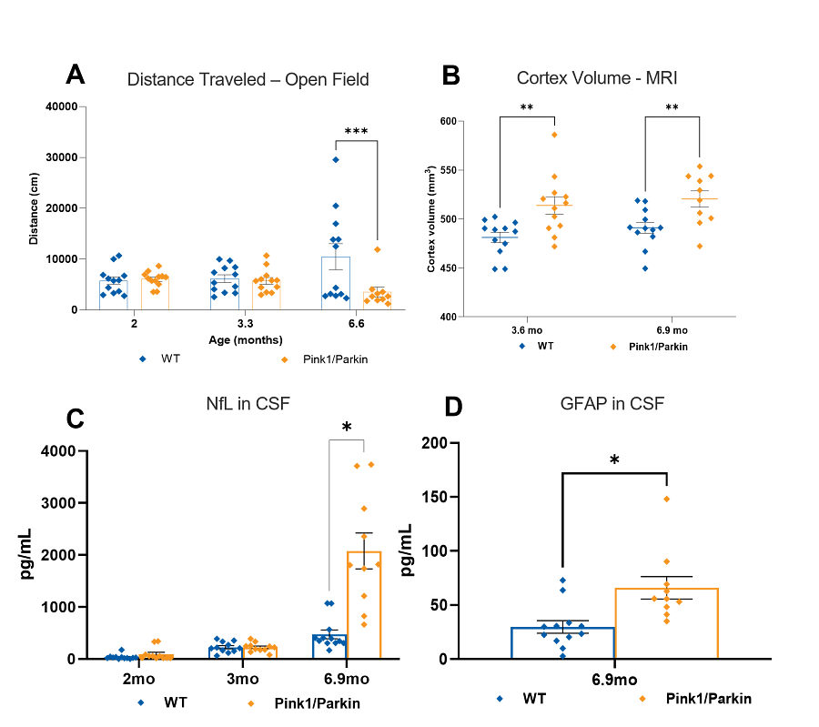
PINK1/PARK2 knockout mice display decreased locomotion and rearing in the open field test at six months of age and have a significantly different gait profile from two months of age when compared to wild-type littermates. Brain volume analysis using MRI reveals increased whole-brain and cortical volume at 6 months of age in PINK1/PARK2 knockout mice. Analysis of biomarkers shows that PINK1/PARK2 knockout mice have increased NfL in plasma from three months of age and CSF from seven months, indicating neuronal damage, and increased GFAP in CSF from seven months of age, indicating astrogliosis.
Alpha-Synuclein Preformed Fibril (PFF) Seeding Model
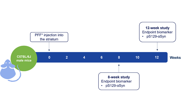
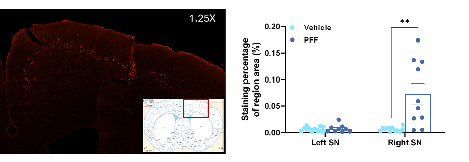
Synthetic alpha-synuclein pre-formed fibrils (PFF) directly injected into the striatum produce a mouse model of alpha-synuclein pathology, by seeding and propagation. Injection of alpha-synuclein PFFs leads to aggregation of phosphorylated S129 alpha-synuclein throughout the mouse brain.
Why choose us for your Parkinson's disease research studies?

Expert Guidance
Our neuroscience experts can guide your model and readout selection, and study design.

End-to-End Services
Comprehensive services from screening to IND-submission support your programs from beginning to end.
Exploratory Toxicology for Neuroscience Drug Discovery
This eBook describes strategies across the early stages of drug discovery to support confidence in your lead small molecule candidate and ensure you proceed through the drug development process with the most promising candidate.
Read the eBook
Frequently Asked Questions (FAQs) for Animal Models of Parkinson’s Disease
-
What animal study models are available for in vivo pharmacology studies of Parkinson's disease?
Charles River utilizes animal models with the well-established neurotoxins 6-OHDA and MPTP. A53T is a familial form of PD, which is also offered as a transgenic model.
-
What are important considerations when choosing the right animal model for Parkinson’s disease research?
A given model should never be used indiscriminately to investigate every question but should instead be carefully selected on the basis of being the most suitable model for the question being asked.