Featured News
Evaluating iPSC-derived Microglia Cell Lines
A reliable tool to assess the efficacy of prospective drugs for neurological diseases in vivo
Microglia are the primary immune effector cells of the central nervous system, and they play a vital role in chronic and acute neuroinflammation. Inflammatory processes and microglial activation in the brain have been linked to a wide range of neurological disorders, including Alzheimer’s disease, Parkinson’s disease, ALS, multiple sclerosis, traumatic brain injury, and more recently, psychiatric disorders including depression. For this reason, there is increasing interest and exploration of neuroinflammatory processes and microglia as a therapeutic target in neuroscience drug discovery.
To fully understand the role of microglia in diverse diseases, and to facilitate effective compound screening to discover new neuroscience therapies, it is essential to establish appropriate cellular models. Microglia isolated from control or disease post-mortem human brain tissue have been used for this purpose. However, the obvious limitations of human brain collection and poor yield of isolated microglia restricts the use of these cells in larger scale compound screening and target validation studies. Microglia derived from induced pluripotent stem cells (iPSCs) may provide an alternative source of cells for such studies.
To examine if iPSC-derived microglia can recapitulate the biological processes of the human brain, scientists from Charles River Leiden compared morphological and functional properties of primary microglial, isolated from post-mortem human brain tissue from the Netherlands Brain Bank, and ioMicroglia™ from bit.bio.
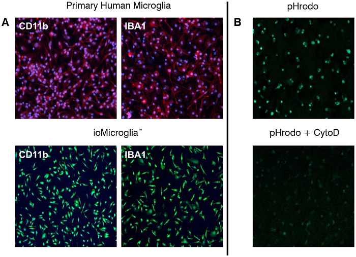
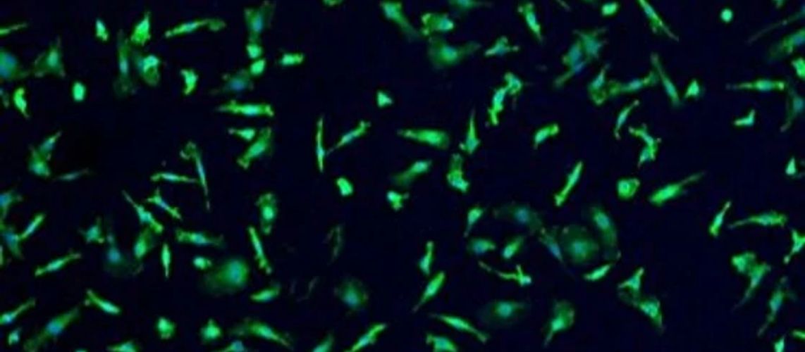
Phenotypic characterization of both cell types shows that human primary microglia and ioMicroglia express key microglial markers such as CD11b, IBA1 TMEM119, and P2RY12. Exposure of primary and iPSC-derived microglia to LPS priming results in increased cytokine secretion in a concentration and time-dependent manner, that could be strongly inhibited by dexamethasone. Concurrent priming and nigericin treatment induced IL-1β and IL-18 release in both cell lines, which was inhibited by the inflammasome inhibitor MCC950. Phagocytosis was investigated in iPSC-derived microglia and human primary microglia by time lapse high-content imaging of the uptake of pHrodo™ Green E. coli BioParticles, and labelled myelin. Time-dependent uptake was observed and was sensitive to disruption of the actin-cytoskeleton with cytochalasin D. In addition, the RNA sequencing data showed that the top 50 expressed genes in primary microglia have similar levels in ioMicroglia, although some differences do exist.
Taken together, these data demonstrated that primary human microglia and iPSC-derived microglia respond similarly to various stimuli and exhibit physiologically relevant processes, such as cytokine release and phagocytosis. Therefore, these cell types could serve as a reliable tool for evaluating the efficacy of prospective drugs for neurological diseases associated with microglia activation and neuroinflammation.