
Overview of Traumatic Brain Injury Models
Traumatic brain injury (TBI) by contusion/impact is the gold-standard model in which novel therapeutics can be tested. We conduct proof-of-concept and efficacy studies in TBI models for acute and chronic therapeutics across multiple modalities, including small molecules, biologicals (antibodies, proteins, etc.), and RNA therapeutics and cell therapies.
Traumatic Brain Injury Research Studies
Alongside appropriate animal models of traumatic brain injury, combinations of readouts across functional assessments, behavior and cognition, preclinical imaging, and bioanalysis/biomarkers enable greater understanding of drug efficacy and effects on pathology. Repeated in-life readouts allow for longitudinal tracking of injury progression and therapeutic efficacy. Our neuroscience experts can advise on the most appropriate combination of readouts to answer your drug discovery questions.
Components for your spinal cord injury study include:
- Post-operative monitoring and assessment as standard
- Behavior tests for locomotor changes and cognitive function
- Fine kinematic gait analysis enabling high-sensitivity detection of subtle changes in gait and limb placement
- Magnetic resonance imaging to assess lesion volume, tissue viability, edema, and blood-brain barrier leakage
- Ex vivo assessment of lesion volume, pathology, and disease biomarkers
Phenotyping of TBI Animal Models
Traumatic brain injury can be induced in rats or mice by mechanical contusion using the PinPoint Precision Cortical Impactor™. This provides precise control over velocity, depth, and power of impact, generating accurate, reliable, and reproducible animal models of traumatic brain injury with a predictable recovery pattern.
-
Injury Progression, Behavior, and Locomotion
Changes in behavior, cognition, and locomotion following traumatic brain injury can be assessed in various ways. Either 7-point or 20-point neuroscore assessments profile cognitive and locomotor deficits can be repeated at multiple time points to assess the effects of acute injury, as well as the impact of therapeutics on recovery. Gross changes in locomotor function can be investigated with limb placement tests or cylinder walking tests. To detect subtle changes in limb movements and gait, kinematic gait analysis can be used.
The data below show the profiling of rats following traumatic brain injury using 7- and 20-point neuroscore assessments. Surgical injury induces an immediate deficit that recovers over three to four weeks.

-
Imaging of Traumatic Brain Injury
High-resolution MRI enables tracking of TBI lesions over time and investigation of therapeutic effects of novel treatments. Beyond simple changes in lesion volume, diffusion-weighted MRI detects changes in cerebral perfusion and can assess the extent of edema in the lesion area. Similarly, MRI with gadolinium contrast agent can assess blood-brain barrier (BBB) leakage and integrity.
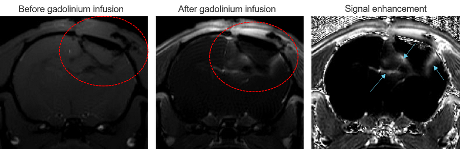
The above images show example MRI images before and after gadolinium contrast injection. The traumatic brain injury lesion is circled in red, and areas of blood-brain barrier leakage are indicated by blue arrows.
-
Histology and Biomarkers
Injury pathology can be assessed through histology and immunohistochemistry, with readouts including lesion size and neuronal cell count via NeuN staining. Similarly, immunohistochemistry enables profiling of neuroinflammatory markers, showing presence of microglia following TBI.
Why Choose Charles River for TBI Studies?

Expertise
Extensive expertise in efficacy study design for brain injury drug discovery

Experience
Over 15 years of experience working with surgical and pharmacological TBI animal models

End-to-End Services
From high-throughput screening to IND-enabling studies

Translational Readouts
Preclinical imaging technologies reflect clinical assessments
This eBook describes strategies across the early stages of drug discovery to support confidence in your lead small molecule candidate and ensure you proceed through the drug development process with the most promising candidate.
Read the eBook