
Surgical CNS Delivery Methods
The blood-brain barrier is a highly selective, semi-permeable membrane that regulates the transfer of substances, solutes, and chemicals between the circulatory system and the central nervous system. It allows the passage of nutrients, such as amino acids, glucose, and nucleosides, as well as diffusion of oxygen, but prevents the passage of large molecules, like antibodies, proteins, and gene therapies. Therefore, specialized CNS drug delivery methods must be used to get large therapeutics to their site of action in the brain. Small molecules are generally administered by standard peripheral methods such as intraperitoneal (IP), intramuscular (IM), or subcutaneous injection.
Our neuroscience team is experienced in a range of CNS drug delivery methods and can provide advice on the most appropriate method for your drug modality. The delivery methods listed below are highly translatable, ensuring appropriate modeling of clinical drug administration:
- Intracerebroventricular (ICV) – injection into the cerebral ventricles
- Intraparenchymal – injection into the brain parenchyma
- Intrathecal – injection into the spinal cord or subarachnoid space
- Nerve bundle administration – injection directly into a nerve bundle, such as the dorsal root ganglion
- Cisterna magna administration – injection into the cerebellomedullary subarachnoid cistern
Validated Cellular & Animal Models for Neuroscience
Our neuroscientists are experts at working with a wide range of cellular and animal models for neuroscience drug discovery, including those for neurodegeneration, neuroinflammation, psychiatric disorders, pain, brain injury, and rare diseases.
Explore CNS models
Following delivery of a therapeutic into the brain, it is important to assess distribution to ensure action in the required brain area, and to investigate potential off-target side effects brought about by action in other brain areas. This can be done a number of ways as part of your drug efficacy studies, including ex vivo methods following sample collection (tissue homogenates, tissue sections, CSF, plasma, or dialysate collected via microdialysis), as well as in vivo imaging methods.
Example methods for the detection of advanced therapies following CNS delivery:
- Detection of viral genome and/or transgene expression with qPCR, ddPCR, hybridization-based branched DNA (bDNA) technology (QuantiGene), or in situ hybridization
- Detection of transgene proteins with immunoassays such as ELISA, MSD, and IHC
- Detection of ASOs with qPCR or hybridization ELISA
- Detection of antibodies by immunoassay
- Detection of antibodies or ASOs by LC-MS following microdialysis
- In vivo nuclear imaging of radiolabeled therapeutic articles
MRI-guided CNS Drug Delivery
A recent major breakthrough in CNS drug delivery methods has been the real-time visualization of gene and cell therapy delivery into the brain. Using real-time, intraoperative MRI (iMRI), coupled with convection enhanced delivery, our scientists are able to deliver advanced therapies to different brain regions with submillimetric (< 0.5 mm) accuracy, reducing off-target effects such as seizures, headaches, fatigue, or tremors. By coupling the test item with a contrast agent, visualized delivery allows us to adapt to ensure accuracy and on-target coverage, bringing your test item where it needs to be.
-
The ClearPoint® System
The ClearPoint® System combines specialized MRI-compatible hardware and software to plan trajectories, and align and guide the infusion cannula to the target area. The cannulas and insertion approach are specifically designed to minimize backflow. Dose volume, infusion rates, and targeting can be optimized throughout this minimally invasive neurosurgery to improve tissue coverage and therapeutic efficacy. The ClearPoint® System is already in use in the clinical environment providing the translational functionality necessary to bridge preclinical safety pharmacology and toxicology with clinical settings
Example Target Sites
Infusion Volume (µL)
Infusion Rates (µL/hr)
Putamen
50 – 250*
120 - 300
Globus pallidus
50 - 125
120 - 300
Thalamus
50 - 125
120 - 300
Caudate Nucleus
20 - 100
120 - 300
Dentate Nucleus
50 - 100
120 - 300
Substantia nigra
10
60
4th Ventricle
400
300
*Up to 200 µL for single site infusion and up to 250 µL (2 x 125 µL) for two infusion sites.
Oblique view of a probe delivering an
AAV-packaged gene to the mid-brain.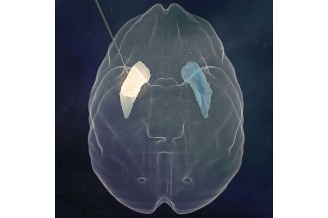
Top view of a probe delivering an
AAV-packaged gene to the mid-brain.