
Using iPSC-Derived Neurons
CNS cells derived from human iPSCs have recently emerged as a straightforward, efficient, and replicable in vitro system for assessing aspects of human neuronal function such as neurite outgrowth. These cultures can be derived from healthy or patient-derived iPSCs. They can be studied in monoculture or co-culture to explore single cell mechanisms as well as the influence of neighboring cell types and signaling interplay. This iPSC technology is ideal for mechanistic studies and disease modeling in human cells.
Methodology to Assess Label-free Neurite Dynamics
Human iPSCs are terminally differentiated into neurons using a specific neuronal differentiation protocol. The effect of a therapeutic compound or inflammatory stimuli on cell viability and neurite outgrowth dynamic are assessed by NeuroTracker software.

Neurites are critical for communication between neurons. Neurodegeneration seen in neuropathological disorders can be attributed to a loss of this connectivity. Neurite outgrowth, complexity, and dynamics influence network connectivity, number of synapses, cell viability, and response to inflammatory stimuli.
iPSC Neurite Tracking
Neuronal differentiation was confirmed by the presence of MAP2 and absence of precursor markers. Neurons are imaged live and the total cell number (yellow) and neurite dynamics (pink) are assessed by NeuroTracker software. Live cell imaging was performed using the IncuCyte® S3 platform.
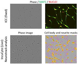
The ability to measure dynamic changes in neurites gives information about the viability of these connections in a healthy or pathological context and the influence of therapeutics on this process.
Studying patient-derived IPSCs can identify drug candidate responders and non-responders to provide a valuable preclinical readout. Overall, they fill an unmet need that bridges the gap between clinical and animal models to support drug development.
Immature Rat Primary Cortical Culture Model
Immature rat primary cortical culture model is a great tool to examine optimal conditions for maximal neurite outgrowth for your neurotherapeutic research. The cortical mixed cultures are prepared from embryonic day 18 rat embryos cortical tissues. On day 3 the exposure of immature cortical culture will be started. Exposure can last for short stimulation periods (10 min) or longer timeframes (up to 72 hours). This allows for a wide treatment window depending on experimental study design for transient pre-treatment, main treatment and for post-treatment.
Studying neurite growth in rat cortical primary neurons makes it possible to identify effective neurotherapeutics, which stimulate and initiate a cortical neuron growth response. This is important to understand in in vitro conditions prior to moving forward to with an in vivo disease model.
Methodology to Assess MAP2 Neurite Dynamics
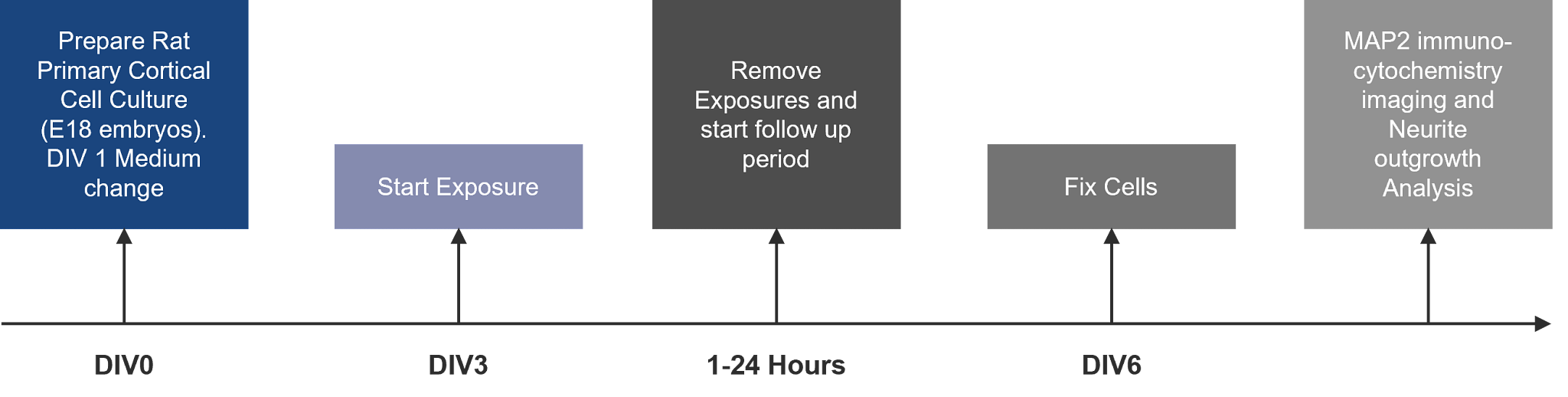
Rat Primary Cortical Neuron Neurite Tracking
Neuronal outgrowth can be quantified by immunostaining the neuronal microtubules with the specific antibody MAP2. The MAP2 protein is involved in the stabilization of the microtubule and intermediate filament complexes located in the neurites of the neurons. Thus, MAP2 is a marker for neurites in neuronal cultures, which are fixed and stained with anti-MAP2 antibody.
The cellular nuclei are stained by adding DAPI (2-(4-Amidinophenyl)-6-indolecarbamidine dihydrochloride), which is cell permeable fluorescent probe for DNA. These will be analyzed by Columbus software by an experimenter blind to the treatments.
Neurite Outgrowth Evaluation Includes:
- Mean total length of processes and branches per cell
- Mean process number per cell
- Mean process and branch number per cell
- Mean branch number per cell
Sholl Analysis
Sholl analysis is a method to measure the dendritic arborization of neurons by maximum number of crossings. The effect of drug treatment on maximum number of neurite crossings can be useful in understanding the efficacy of your candidate.
Automated rat cortical neurite outgrowth analysis using CSIRO algorithm
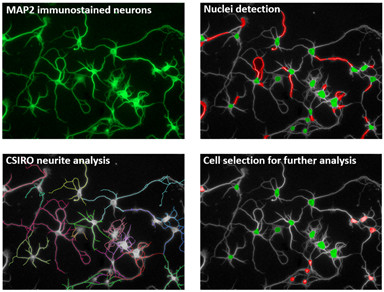
Neurite Outgrowth Assay and Quantification
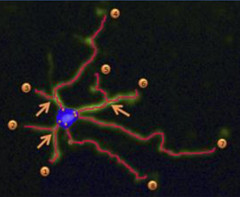
The image shows automated neurite outgrowth analysis using the CSIRO algorithm for quantification of maximum neurite length, number of extremities, number of roots, number of nodes, and total neurite length.
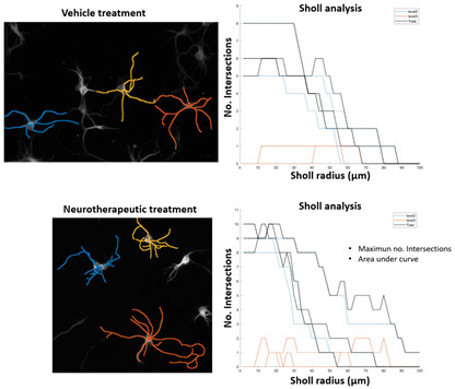
Sholl Analysis measures the dendritic arborization of MAP2 stained rat cortical neurons. The maximum number of intersections and Area under curve (AUC).
The ability to measure dynamic changes in neurites gives information about the viability of these connections in a healthy or pathological context and the influence of therapeutics on this process.
Frequently Asked Questions (FAQs) for Neurite Outgrowth Assays
-
What is neurite outgrowth?
Neurite outgrowth is a process where developing neurons produce new projections as they grow in response to nerve growth factors or neurotrophins.
-
When does neurite outgrowth become compromised?
Aging is one of the leading causes of neuron atrophy; however, brain lesions that occur with acute brain injuries or with chronic inflammation from neurodegeneration also result in impaired sprouting/decreased neurite outgrowth.
-
Can non-neuronal cells in the brain influence neurite outgrowth?
Diminishing glial fibrillary acidic protein (GFAP) levels of old astrocytes or compromised astrocytes due to excess inflammation, can affect neurite outgrowth. Cellular senescence increases with age and several laboratories suggest that both aging and age-related neurodegenerative diseases are accompanied by an increase in senescent cells of non-neuronal origin in the brain.