Functional Assays in Isolated Microglia
Microglia are the resident immune-effector cells in the central nervous system and amount to 10-15% of all brain cells. Microglial activation typically occurs in response to brain injury, neurodegeneration, and infection, resulting in altered function and the release of inflammatory mediators which may be beneficial or detrimental, depending on the microenvironmental context.
Microglia are highly sensitive to changes in their environment. This is particularly evident during isolation and culturing of microglia in vitro with serum. Although numerous methods of isolating intact microglia are widely used, the majority lead to cells that are both highly proliferative and amoeboid, hallmarks of microglial activation. At Charles River we have adopted culturing methods to better recapitulate the morphology and phenotype of ‘resting’ microglia, enabling improved modelling of the in vivo environment in functional assays.
A battery of functional microglial assays, including cytokine production, inflammasome activation, and microglia phagocytosis assays, can be used to examine microglial activation, and to assess whether immunomodulating therapeutics can dampen or enhance microglial activation in vitro. The assays presented below can be conducted in primary mouse microglia (wild-type and disease models), human post-mortem isolated microglia, as well as human iPSC-derived microglia. Microglial function assays can also be combined with neuronal function assays, such as micro-electrode arrays (MEA) in neuron-glia co-cultures.
Technical Sheet: In Vitro Microglial Assays
Learn more about our functional assays to investigate the effect of novel therapeutics on microglial phagocytosis, inflammasome activation, and cytokine release profiles.
Download now
Isolation and Purification of Mouse Microglia
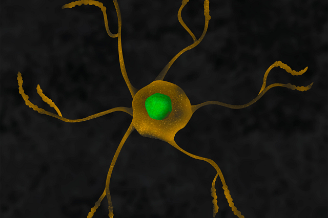
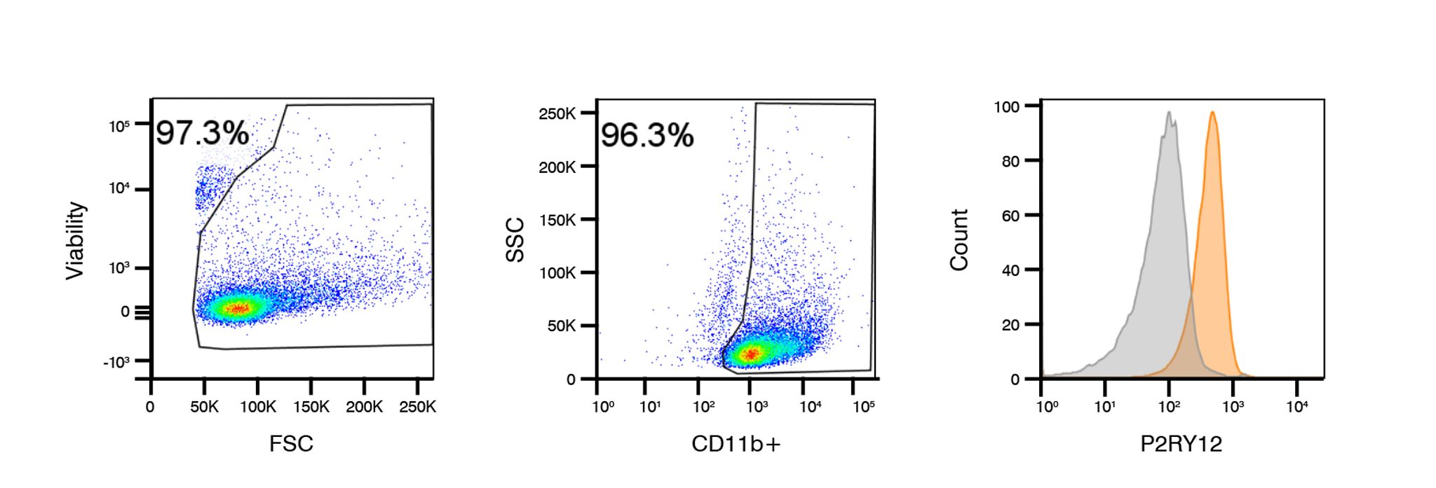
Adult mice brains are dissected and dissociated into single cells, and subsequently sorted by magnetic selection and purity confirmed by CD11b/P2RY12 expression using flow cytometry. Purified mouse microglia are cultured for 6 days in conditioned culture medium supplemented with defined astrocytic factors. Cultured mouse microglia exhibit a ramified small-bodied morphology turning amoeboid during microglial activation, following LPS stimulation. Images below were taken using the IncuCyte® S3 platform.
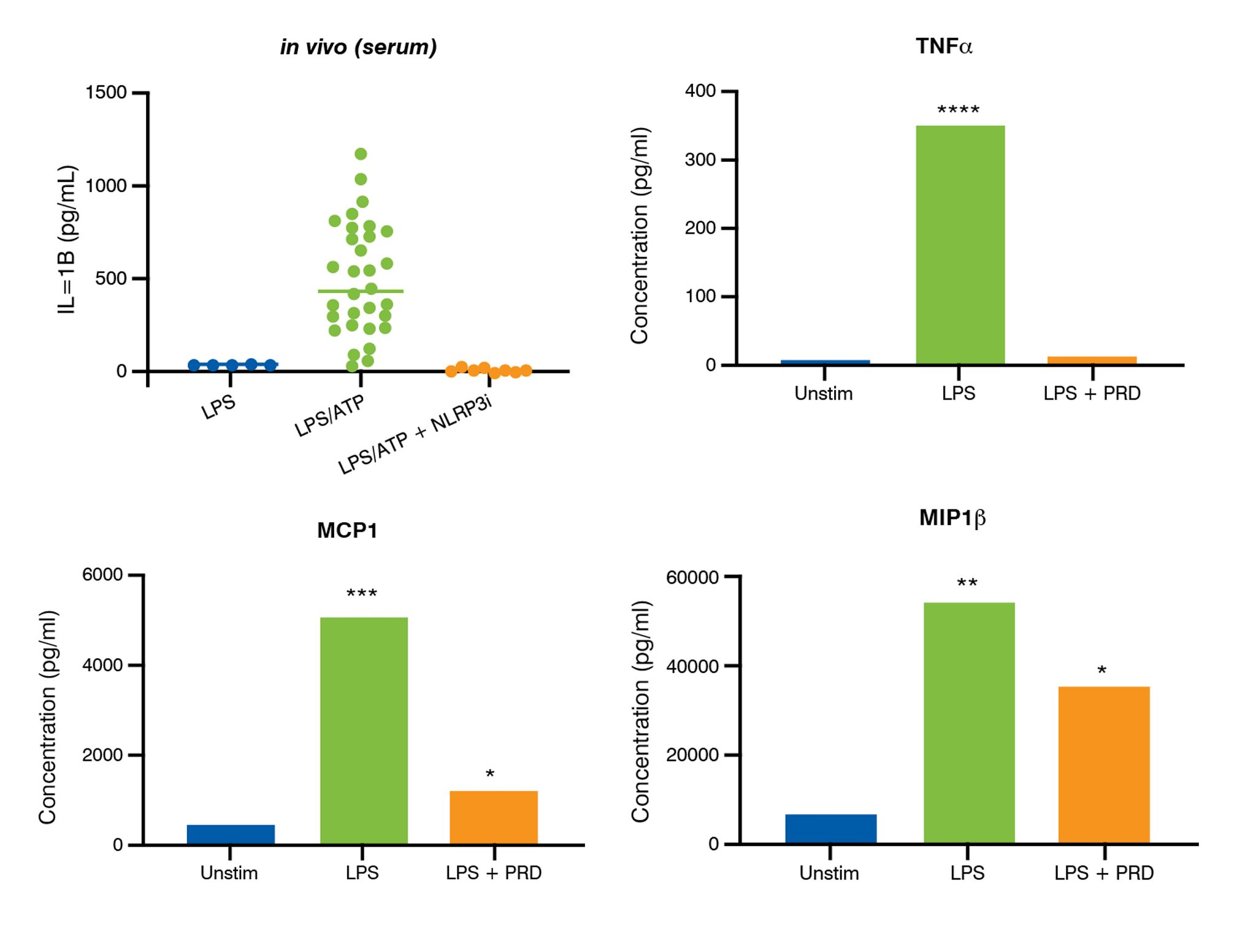
Microglia isolated from adult mice are highly pure, express microglia phenotypic markers, and adopt a ramified morphology in defined culture conditions. Cultured mouse microglia release pro-inflammatory cytokines upon stimulation with LPS that can be inhibited using corticosteroids (e.g., prednisolone, PRD).
Microglial Phagocytosis Assays
Phagocytosis, in response to distinct molecular signals, is a key function of microglia required to maintain homeostasis and is a key readout of their normal function. Therapeutic manipulation of this function can drive the clearance of aberrant disease material such as dysfunctional cells, or aggregated proteins such as α-Synuclein and β-amyloid. Similarly, in some disease circumstances, reducing overactive phagocytic clearance may be therapeutically beneficial.
Adult mouse microglia cultured under serum-free conditions efficiently phagocytose bio-particles and their phagocytic function can be either enhanced or suppressed depending on the mechanistic intervention. In vitro screening utilizing adult microglia derived from healthy or disease model tissue can provide information on mode of action and impact on cell function of therapeutics targeting microglial cells.
In Vitro Microglia Phagocytosis Assay Methodology
Microglia are purified from adult mouse brains and pre-incubated with the therapeutic substance. Target materials (e.g., beads or bio-particles) are labelled and applied to cultured microglia, following activation with LPS. Phagocytosis is then kinetically monitored in real-time over 24 hours using the IncuCyte S3 platform, with the rate of uptake analyzed. The in vitro microglia phagocytosis assay can be used to investigate the effect of novel therapeutic on microglial phagocytosis of disease relevant materials, such as amyloid-β species.
Microglial Inflammasome Activation Assays
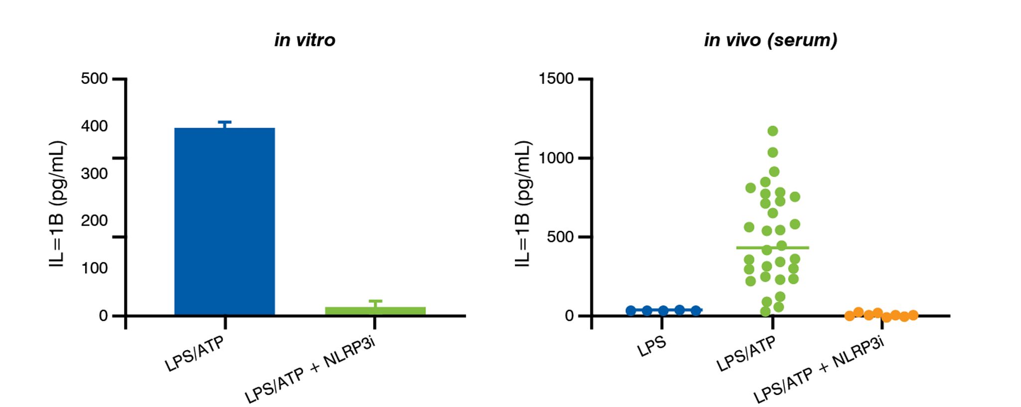
Activation of the NLRP3 inflammasome in microglial has been implicated in the pathophysiology of a variety of disorders with a neuroinflammatory component. Measurement of inflammasome activation in in vitro microglial assays enables compound selectivity and efficacy against inflammasomes to be determined prior to translation into in vivo studies.
Microglial Cytokine and Chemokine Assays
Under physiological conditions, activated microglia release pro-inflammatory cytokines and chemokines, and this is recapitulated through activation with LPS in cultured microglia. Measurement of cytokines and chemokines from activated microglia can be achieved through Bio-Plex Multiplex immunoassays (Luminex®). Supernatants are collected following incubation of microglia with test compounds to assess therapeutic efficacy.
Frequently Asked Questions (FAQs) for Mouse Microglial Activation Assays
-
What are the benefits of using adult microglia over more commonly used neonatal cells?
Current microglial models of neuroinflammation include microglial cell lines or primary microglia prepared from embryonic or neonatal mice. Increasingly, studies report diverse microglial phenotypes that are linked to development showing distinct molecular signatures. Commonly studied microglia cell lines and embryonic or neonatal-derived microglia show differing responses to stimuli and express strikingly different molecular signatures compared to adult microglia. The use of adult microglia is especially pertinent in the field of neurodegenerative diseases where aging is a critical parameter.
-
How can IncuCyte® Phagocytosis Assays help me to test my therapeutic?
Measuring phagocytosis in real-time using the IncuCyte system allows you to visualize and quantify microglial phagocytosis activity over time, adding a higher level of mechanistic insight which can complement endpoint phagocytosis readouts by methods such as flow cytometry or immunofluorescence. A plethora of target materials such as synthetic beads, protein aggregates, or cells can be labelled with high specificity allowing a highly-tailored assay approach. This method can be readily scaled up to test several compounds in response to different stimuli in comparison to a positive or negative control drug. Together, this assay offers a flexible, bespoke platform to test microglial function which can be tailored to your therapeutic and target of interest.
-
Could these functional microglial assays be applied to other in vitro microglial models?
Yes – while the data above are using isolated mouse microglia, these assays could also be used to investigate the effect of a novel therapeutic in stem cell derived microglia or post-mortem isolated human microglia.