
3D Cell Culture Models
90% of drugs that seem promising in vivo ultimately fail in the clinic. Collecting more predictive, translational data that represents a therapy’s interaction with the human immune system and tumor microenvironment (TME) is essential to predicting clinical success, and yet not all programs take this approach.
Optimal Translation: 3D Tumor Models
In conjunction with our drug sensitivity and molecular characterization data, our 3D tumor models allow you to evaluate the potency of your compound compared to standard-of-care drugs. Created using our proprietary patient-derived xenograft (PDX Model) explants or cell line-derived xenografts (CDX models) the 3D tumor models allow you to identify the most promising drug candidates, tumor histotypes, and molecular subtypes for further testing.
Our 3D cell culture models can support you oncology and immuno-oncology drug development and includes a range of immune cell-mediated tumor killing assays.
Our 3D tumor models and services include:
- Clonogenic Assay 3D tumor models
- Combination studies
- Integration with in vitro and in vivo PDX models

White Paper: How PDX In Vitro Assays Are Accelerating Oncology Developability
This white paper describes that by harnessing PDX advantages with next-generation tools such as 3D technology and machine learning live cell imaging we can pave the way for a quicker path to clinical application.
Download the White Paper
Clonogenic Assay 3D Tumor Models
In the clonogenic assay, patient-derived tumor samples or tumor cell lines are cultured to form ex vivo 3D tumor organoids. Compound effects are then evaluated using image analysis.
Clonogenic Assay 3D Tumor Models
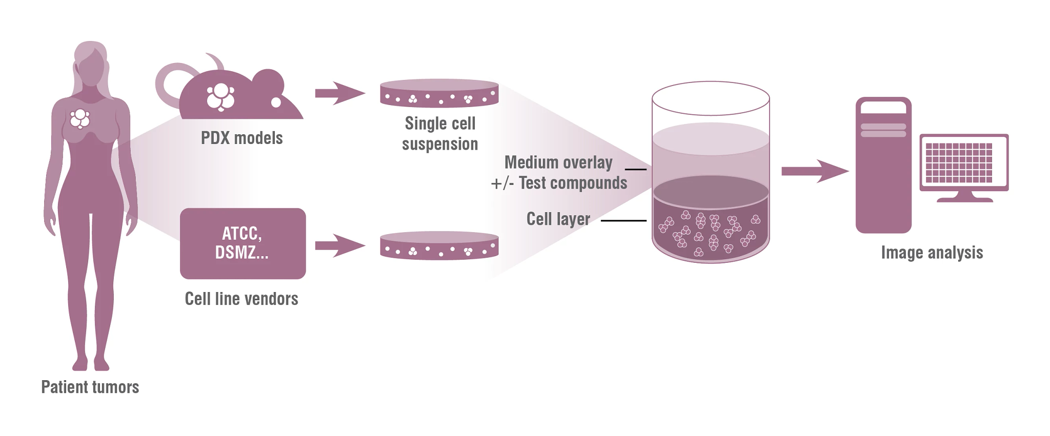
Figure 1: Schematic of ex vivo 3D tumor models in Clonogenic Assay format.
Tumor panel screens in 3D cell culture models with PDX or CDX cell suspensions can be used to assess the efficacy of oncology therapies to inhibit anchorage-independent growth of single cells and ex vivo formation of tumor organoids in single agent efficacy or matrix combination layouts. These 3D tumor models can investigate if anti-tumor activity is selective for different tumor histologies or dependent on molecular markers, allowing therapeutic indications to be determined, and indicate candidate tumor models for subsequent in vivo studies.
Download our 3D Tumor Clonogenic Assay Data Sheet for more information.

Patient-Derived Xenografts – Cancer Model Database
Support your in vitro, in vivo, and ex vivo studies with a user-friendly search, new model data (including HLA typing, growth curves, and tumor images), and multi-parameter search options for all tumor model types (PDX and CDX).
Visit our Database
PDX Models and Combination Studies
3D assays can be used to run systematic two-drug combination therapy studies over a range of concentrations and to correlate the tumor response with molecular data (biomarker identification). Data collected in 3D tumor models can then be used to select PDX models for subsequent in vivo efficacy tests.
Integrating In Vitro and In Vivo PDX Models
Our fully integrated approach using in vitro 2D and ex vivo 3D tumor models, including immune cell mediated tumor killing assays with in vivo PDX models and bioinformatics data will facilitate drug development and enhance the speed of your oncology research.
Frequently Asked Questions (FAQs) About 3D Tumor Models
-
What is a patient-derived xenograft?
Patient-derived xenografts (PDX) are models of cancer where the tissue or cells from a patient’s tumor are implanted into an immune deficient or humanized mouse. PDX models simulate human tumor biology allowing for natural cancer progression, and offer the most translational research model for evaluating efficacy. Determine which PDX model is best for your studies
-
Does in vitro data correlate with in vivo PDX models?
Data collected in in vitro 3D tumor models can be used to select PDX models for subsequent in vivo efficacy studies. Learn about the data correlation in our article ‘A High-Content Image Analysis Approach for Quantitative Measurements of Chemosensitivity in Patient-Derived Tumor Microtissues’
-
What is a clonogenic assay?
Clonogenic assays are a type of 3D tumor models can be used to determine cell reproductive death after treatment with radiation and other cytotoxic agents. This technique allows you to study the effectiveness of specific agents on the survival and proliferation of cells.
Learn More about Clonogenic Assays -
What oncology models are right for me?
In a complex and changing field, knowing the next step can be difficult. Answer a few brief questions about your research to receive expert guidance on how to advance your oncology program; from 3D tumor cell culture models to in vivo pharmacology studies.
Receive Personalized Advice