
Cytotoxic T Cell-Based Tumor Killing Assays
Charles River have a range of immune mediated tumor killing assays including cytotoxic T cell assays and natural killer (NK) cell assays. These assays are analysed using flow cytometry and live cell imaging (IncuCyte); IncuCyte-based assays quantify a target number that can be multiplexed with target specific apoptotic readout, and can be tailored to the anticipated mechanism of action by selecting from a panel of validated target cell lines.
Immune Mediated Tumor Killing Assays
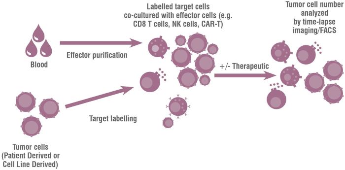
Figure 1: Diagram of Immune Mediated Tumor Killing Assays used to evaluate oncology immunotherapies.
Our panel of immune-mediated tumor killing assays include:
- IncuCyte® tumor killing assays (2D)
- Macrophage-dependent ADCC assays
- IncuCyte® spheroid tumor killing assays (3D)/CDC assays
- Flow cytometry-based killing assays
- Natural killer (NK) cell tumor killing assays
Which target cell lines are validated for tumor killing assays?
Charles River has an extensive list of 2D and 3D validated tumor cell targets from a range of tissue types including lung, ovarian, pancreatic and intestines. Where a specific line is required, cells are transduced with a nuclear RFP and a pilot experiment is run to determine the optimal seeding densities ahead of efficacy/mechanistic testing. This process has also been successfully applied to PDX cell lines after identifying relevant gene expression using our cancer model database.

Cancer Model Database
Support your in vitro, in vivo, and ex vivo studies with a user-friendly search, new model data (including HLA typing, growth curves, and tumor images), and multi-parameter search options for all tumor model types (PDX and CDX).
Visit our Database
IncuCyte tumor killing assays (2D)
Cytotoxic T cells can be cultured with activating antibodies such as anti-CD3 and tested for their ability to kill tumor targets in an IncuCyte-based assay. The ability of novel therapeutics to potentiate cytotoxic T cell killing can then be assessed by quantifying the change in the number of viable tumor cells over time. Positive control agents which enhance tumor killing have been validated in this assay. Caspase 3/7-dependent tumor cell apoptosis can also be determined by co-localization of the caspase signal to NucLight Red positive tumor cells. The target cell population can be adapted to suit the disease indication or mechanism of action.
Data: Tumor killing assays (2D)
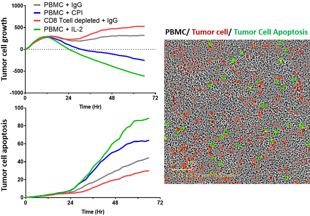
Figure 2: PBMC - mediated tumor killing in 2D format, and example image, demonstrating killing over time with checkpoint inhibitor treatment with or without depletion of CD8+ cytotoxic T cells. Alt tag: PBMC - mediated tumor killing assay in 2D format, demonstrating killing over time with checkpoint inhibitor treatment with or without depletion of CD8+ cytotoxic T cells.
IncuCyte spheroid immune mediated PDX tumor killing assays (3D)/CDC assays
Our team can also run immune cell killing assays with PDX (patient-derived xenograft) material; in vitro 3D assays incorporate live cell imaging to predict in vivo responses to matched PDX material. Screening PDX models in vitro prior to in vivo humanized tumor killing models enables stratification and prioritization of the wide range of in vivo PDX models available.
Data: Tumor killing assay (3D)
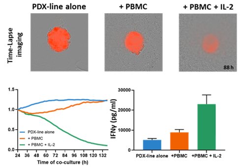
Figure 3: PBMC - mediated tumor killing in 3D format, and example images, demonstrating killing over time with or without IL-2 stimulation. Supernatants were harvested and levels of IFNg analysed as a surrogate marker to effector cell activation.
The combination of in vitro screening with in vivo trial design options will aid your drug development and increase throughput.
Have a question or need advice on which assay is right for you?
Frequently Asked Questions (FAQs) About Tumor Killing Assays
-
Can I test my therapeutics in both PBMC and purified cell assays?
Charles River offers both PBMC and purified cell assays. For example, purified CD8 T cells or monocytes can be co-cultured with target cells alone or in combination. These types of studies can help to refine the cellular target and understand which cell types work together to mediate tumor cell killing.
-
Can I use murine lines to predict in vivo efficacy in tumor models?
Murine tumor cell lines are compatible with nuclear RFP transduction and we see variation in the extent of killing depending on the strain of murine PBMC used in co-culture. Murine tumor killing assays are useful to refine test conditions ahead of limited and expensive in vivo studies.
-
How do I know my target is expressed on the tumor cell lines you commonly use?
Our team can screen cell lines for your target expression by flow cytometry, western blot or qPCR. Target cells can also be induced to express a target, for example, IFNγ stimulates upregulation of PD-L1 by many tumor lines. Alternatively, genetic manipulation can force expression or deletion of required proteins.
-
Is it better to test our therapeutics in 2D or 3D PBMC cell tumor killing assays?
This depends on your target and therapeutic approach. 2D IncuCyte assays enable target and apoptotic readouts while 3D formats better model the in vivo tumor structure. If successful, testing could then progress to in-vitro/in-vivo PDX testing or syngeneic mouse models. Our team of immunologists can advise the best assay for your purpose.
-
Is it better to test our therapeutics in flow-based or IncuCyte-based assays?
Flow-based assays are ideal for suspension tumor target cells while the IncuCyte platform is best suited to adherent tumor cell lines. Flow cytometric based assays enable additional mechanistic information via inclusion of surface markers and intracellular cytokine staining, providing the ability to assign function to specific PMBC cell subsets, which is not possible using the IncuCyte. For example, expression of activation markers (such as CD25 and CD69) or effector molecules (e.g., IFNγ and Granzyme B) by phenotypically distinct PBMC subsets can be quantified alongside therapeutic targets or potential inhibitory receptors like PD-1. IncuCyte imaging is adaptable to higher-throughput screening and gives a highly quantitative and visual representation of the ability of your therapeutic to modulate tumor killing. Charles River can work with you to select the best assay for your program.