
Defined Tumor Tissue Microarray Plates to Support Model Selection
Containing >700 tumor models grouped by tumor entity, our current, sixth generation of tumor microarray allows us to individually screen cancer types by selecting from:
- Nine individual patient-derived (PDX) tumor plates (548 models)
- Two cell-line derived (CDX) tumor plates (100 models)
- Complemented by stand-alone brain tumor PDX plate (56 models) launched in January 2023
- One syngeneic tumor plate (32 models)
Syngeneic Model Data
Access our new syngeneic model data including checkpoint inhibitor (e.g. anti-PDL-1, anti-PD-1, anti-CTLA-4), whole exome sequencing (WES), and RNA-seq data. This data gives you the ability to choose based on mutation status in addition to sensitivity immune checkpoint inhibitors.
Download now
Tissue microarray immunohistochemistry (IHC) is most commonly used for identification and confirmation of suitable tumor models by means of target protein expression (Figure 1). Charles River has a range of established antibody staining protocols to visualize key oncology targets, and we can also support development of novel targets.
Alternatively, you can receive our tumor microarrays slides to run your established assays yourself.
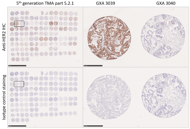
Figure 1. Tissue microarray immunohistochemical staining of tumor microarray plate containing various cancer types (anal, gastric, head and neck, esophageal, liver) to identify HER2-expressing tumor models. GXA 3039 and GXA 3040 are shown as examples for highly expressing and non-expressing tumor models, respectively, alongside the corresponding isotype control staining. Scale bares: 5 µM.
Each tumor model is represented by two individual biopsy cores of 1 mm diameter, sampled from representative regions of the grafted tumor, and arranged side by side on the microarray plates. A Hematoxylin and Eosin (H&E) counterstained digital slide of an early section from each TMA part is available on request as part of our service and allows assessment of the general histology of the biopsies. Upon identification of suitable tumor models by tumor tissue microarray screening on biopsy level, additional analyses can be performed on whole tumor sections for individual models to gain further insights.
Identify the Most Relevant Tumor Microarray for Your Project
Charles River offers a variety of tumor tissue microarray plates to help you select the most appropriate tumor model before proceeding to in vivo tumor models.
-
PDX Tumor Microarray Plates
Histotype Cancer subtypes (abbreviation, no. of models) CRL Reference Anal Anal cancer (AXF, 7) 6.2.1 Bladder Bladder carcinoma (BXF, 10) 6.7.1 Brain Central nervous system cancer (CNXF, 44), Neuroblastoma (NBXF, 12) Brain TMA Breast Of Asian ethnicity (MAXA, 1), Her2-enriched (MAXFHER, 8), Luminal B subtype (MAXFLB, 1), Triple negative (MAXFTN, 15) 6.3.1 Cervix Cervix carcinoma (CEXF, 5) 6.3.1 Colon Of Asian ethnicity (CXA, 5), Other colon cancer (CXF, 74) 6.2.1, 6.1.1 Epidermis Epidermal carcinoma (SKXF, 3) 6.6.1 Esophageal Esophageal cancer (OEXF, 1) 6.2.1 Gastric Of Asian ethnicity (GXA, 29), other gastric cancer (GXF, 7) 6.2.1 Gastrointestinal Gastrointestinal stromal carcinoma (GIXF, 2) 6.6.1 Head & Neck Head and neck cancer (HNXF, 10) 6.2.1 Kidney → See renal Leukemia Acute lymphoblastic (LEXFAL, 1), acute myeloid (LEXFAM, 13) 6.9.1 Liver Cholangiocellular carcinoma (LIXFC, 8), Hepatocellular carcinoma (LIXFH, 2) 6.2.1 Lung Of Asian ethnicity (LXA, 4), NSCLC adenocarcinoma (LXFA, 44), NSCLC of Asian ethnicity (LXAA, 1), other (LXF, 2); NSCLC epidermoid (LXFE, 36), NSCLC large cell (LXFL, 10), SCLC (LXFS, 11) 6.4.1, 6.5.1 Lymphoma Diffuse large B cell (LYXFDLBC, 14), Hodgkin (LYXFH, 1) 6.9.1 Melanoma Melanoma (MEXF, 31) 6.9.1 Ovary Ovarian cancer (OVXF, 18) 6.3.1 Pancreas Pancreatic cancer (PAXF, 50) 6.8.1 Pleuramesothelioma Pleuramesothelioma (PXF, 9) 6.9.1 Prostate Prostate carcinoma (PRXF, 4) 6.3.1 Renal Of Asian ethnicity (RXA, 1), other (RXF, 46) 6.7.1 Sarcoma Of Asian ethnicity (SXA, 24), Ewing (SXFE, 1), Osteosarcoma (SXFO, 4 , Soft tissue sarcoma including rhabdomyosarcoma, myosarcoma, liposarcoma and fibrosarcoma (SXFS, 10) 6.6.1 Skin → See melanoma Thyroid Thyroid cancer (THXF, 4) 6.3.1 Testis Testicular carcinoma (TXF, 2) 6.3.1 Uterus Of Asian ethnicity (UXA, 1), other (UXF, 1) 6.3.1 -
CDX Tumor Microarray Plates
Histotype Cancer subtypes (abbreviation, no. of models) CRL Reference Brain Central nervous system cancer (CNXF, 3) 1.1 Breast Her2-enriched (MAXFHER, 1), Luminal B subtype (MAXFLB, 2), Triple negative (MAXFTN, 2) 1.2 Colon Colon cancer (CXF, 15) 1.1 Gastric Of Asian ethnicity (GXA, 6) 1.2 Kidney → See renal Leukemia Acute lymphoblastic (LEXFAL, 3), acute promyeloic (LEXFAM, 10), chronic lymphatic – B cell (LEXFCL, 1), chronic myeloid (LEXFCM, 5) 1.1 Liver Hepatocelluar carcinoma (LIXFH, 3) 1.2 Lung NSCLC adenocarcinoma (LXFA, 4), NSCLC epidermoid (LXFE, 3), NSCLC large cell (LXFL, 2), SCLC (LXFS, 2) 1.2 Lymphoma Diffuse large B cell (LYXFDLBC, 3), non-Hodgkin centroblastic (LYXFNH, 4) 1.1 Melanoma Melanoma (MEXF, 1) 1.2 Myeloma Multiple (MMXF, 8) 1.1 Ovary Ovarian cancer (OVXF, 8) 1.2 Pancreas Pancreatic cancer (PAXF, 3) 1.2 Prostate Prostate cancer (PRXF, 6) 1.2 Renal Renal cancer (RXF, 3) 1.2 Skin → See melanoma Squamous Squamous carcinoma (VXF, 1) 1.2 Uterus Uterus cancer (UXF, 1) 1.2 -
Syngeneic Tumor Microarray Plates
List of models 11509F #7802, 11509F #7803, 4T1, 4T1_FR, A20, B16F10, BRAC(ex2)-/-, C1498, Cloudman S91, Colon26, CT26, E.G7-OVA, EL4, EMT6/P, F9_FR, GL261, J558, KLN205, KP, KP1, KP4, L1210, Lewis Lung_FR, LL/2, Madison 109, MBT-2, MC38, N1E-115_FR, P388, Panc02 wt_FR, Panc02_7-1-17_FR, Renca_FR
For more information on our tumor models please visit our cancer model database or our oncology models page.