Monocyte Cell Migration Assay
Monocytes are innate immune cells involved in resolving tissue injuries, inflammation, and infections. Monocyte migration is driven by chemokines, including the stromal cell-derived factor 1α (SDF-1α), which is released by residential cells upon tissue damage1-4.
SDF-1α, also known as CXCL12, allows monocytes to migrate through the vascular or lymphatic endothelium to the peripheral injured or inflamed tissue. Here, monocytes can further differentiate into macrophages or dendritic cells5 and signal through the SDF-1α receptor, CXCR4. Although the recruitment of monocytes is essential for mounting effective immune responses and restoring tissue homeostasis, it also plays a pivotal role in the development of several inflammatory disorders and tumor development.
Hence, monitoring the response of human monocytes to chemokines is key for the identification of candidate drugs. To study monocyte migratory mechanisms and test the effects of drug candidates on the modulation of monocytic migration, we have developed a chemotaxis assay using primary human monocytes freshly isolated from blood of healthy donors. Results from these in vitro assays are predictive for in vivo translation.
Assay Principle
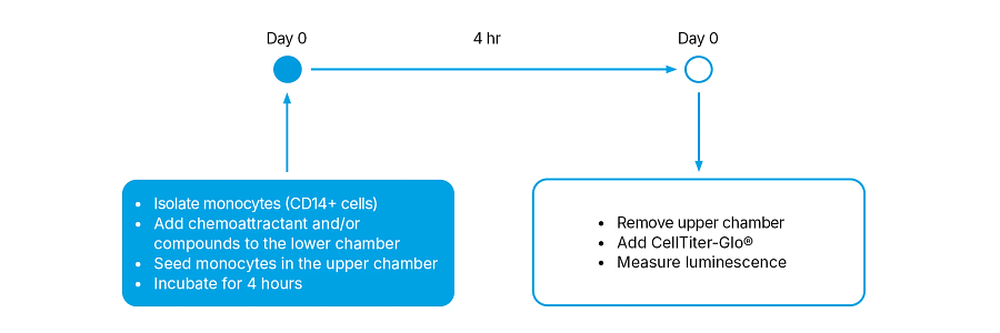
The chemotaxis assay evaluates the migration of monocytes through permeable supports (Transwell® or Boyden chamber) that contain 5.0 μm pore polyester membrane. Blood-derived human monocytes are isolated using magnetic beads coupled to an anti-CD14 antibody (positive selection) and seeded in the upper chamber of a 96-well Boyden chamber in serum-free medium, while chemoattractants and/or compounds are placed in the lower chamber. After 4 hours, monocytes that have migrated through the pores into the lower chamber are detected by measuring their cellular ATP levels using a luminescence-based reaction (CellTiter-Glo®, Promega) and read with a plate reader (EnVision™, PerkinElmer). The luminescent signal generated is directly proportional to the number of cells present in the lower chamber (Fig. 1).
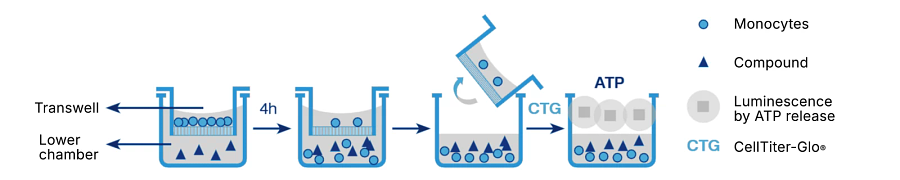
Figure 1. Chemotaxis assay principle with human monocytes
Assay Workflow
Assay Setup
| Cell type | CD14+ cells (monocytes) from healthy human blood donors |
| Seeding density | 50,000 cells/insert |
| Trigger | SDF-1α [10 nM |
| Controls | Control: Serum-free medium (0.5% BSA) Vehicle: 0.1 % DMS0 |
| Inhibitors | AMD3100 (CXCR4 inhibitor) in 8-point concentration-response curve |
| Time | 4 hours |
| Readout | ATP levels (luminescent signal) |
Quality Check (QC) of Monocytes
Flow cytometry is used to perform a quality check (QC) of freshly isolated monocytes. After isolation, monocytes are stained with an anti-CD14 antibody and its relative isotype control, and analyzed by flow cytometry (Fig. 2A). Donors that exhibit > 80% of CD14 expression will be used for the chemotaxis assay (Fig. 2B).
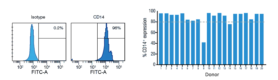
Figure 2. Monocytes (CD14+ cells) quality check
Assay Performance
Performance of the developed assay was validated using the SDF-1α receptor antagonist, AMD3100 (CXCR4 inhibitor), as a positive control for inhibition of migration together with the two assay controls (Control: Serum-free medium [0.5% BSA]; Vehicle: 0.1% DMSO). Representative data obtained from monocytes (CD14+ cells) isolated from two healthy donors (Donor 01 and 02) are reported below (Fig. 3).
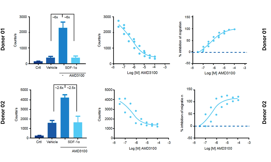
Figure 3. Monocyte chemotaxis responses
Summary
Migration of immune cells is essential for resolving tissue injuries, inflammation, and infections. This process is orchestrated by chemokines, such as SDF-1α, which can recruit monocytes and other immune cells. SDF-1α, a member of the C-X-C chemokine family, has been reported to be implicated in different pathologies including pancreatic cancer6, Alzheimer’s disease7, atherosclerosis8 and Sjögren’s syndrome9. Therefore, a better understanding of the processes involved in migratory responses of monocytes would offer insight into potential treatments for inflammation and autoimmune diseases.
Here we demonstrate an optimized chemotaxis assay allowing the modulation of SDF-1α-induced migration of human monocytes isolated from healthy blood donors through a 5 μm pore size inserts of a Boyden chamber. SDF-1α at 10 nM strongly induced the migration of human monocytes that is inhibited by the CXCR4 antagonist (AMD3100) in a dose- dependent fashion. IC50 values were consistent across different donors (not shown). Using this assay, the migration of monocytes isolated from blood donors can be monitored to evaluate therapeutic candidates for the treatment of inflammatory conditions and other pathologies.
- Complementary Immunology Assays
-
References
- Luster AD, New England Journal of Medicine, 1998, 338, 436-445.
- Sánchez-Martín L et al. Blood, 2011, 117, 88-97.
- Deshmane SL et al. Journal of interferon & cytokine research, 2009, 29, 313-326.
- Shi C & Pamer EG Nature Reviews Immunology, 2011, 11, 762.
- Imhof BA & Aurrand-Lions M, Nature Reviews Immunology, 2004, 4, 432.
- Cui R et al. Oncotarget, 2016, 7(31), 50735.
- Raman D et al. Toxicol Appl Pharmacol. 2011, 256: 300–313.
- Potteaux S et al. J Clin Invest. 2011, 121:2025-36.
- Ushio A. et al. World J Immunol. 2017; 7: 1-8.