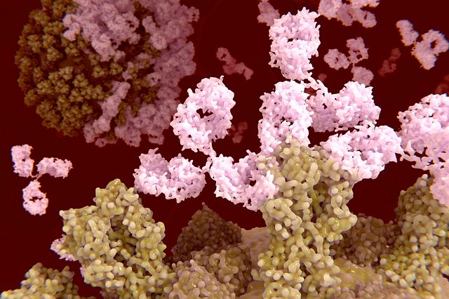
Animal Models of Inflammation and Autoimmune Disease
When our immune system is inappropriately activated this can result in chronic inflammation and autoimmune disease. Advances in immunology research have clarified the roles the immune system plays in many different disease areas, including those that have not been traditionally connected with immunity. Charles River’s expert immunologists can support your immunology programs with a wide selection of in vivo and in vitro immunology assays, inflammation models, and autoimmune models, across numerous therapeutic areas, like vaccine development, infectious disease, and immuno-oncology.
In a truly translational approach to discovery and development, we combine our inflammation models and autoimmune models with relevant biomarkers, like flow cytometry and histologic analysis, for a study design tailored to meet the goals of your immunology programs.

Most probiotic screening skips the immune system.
We don’t. Our in vitro platform reveals how strains modulate human immune cell function, so you can make smarter decisions faster. See how we demonstrate anti-inflammatory effects.
Download the whitepaper
Challenges in Immunology Research
Global immunosuppressive drugs that treat autoimmune and auto-inflammatory disease (cyclosporin A and rapamycin) might soon be replaced by newer immunomodulatory therapies targeting the cell types and pathways that drive the disease process. The advantage of such therapies is that the rest of the immune response remains intact, with better outcomes for patients.
Inflammation and autoimmune drug discovery programs that focus on a specific disease area require models that recapitulate the human target disease as closely as possible (such as autoimmune and inflammation models). It is important to choose the best efficacy model to elucidate the effect of your therapeutic. For example, if your target is CD4 Th1 T cells, your chosen autoimmune disease model should demonstrate a strong Th1 component.
Physiological Relevance Shouldn’t Be Optional.
Standard T cell assays lack the nuance needed for cancer vaccine and IO drug development. Our platform brings you closer to clinical outcomes with real immune specificity. Explore the new gold standard.
Download the whitepaper
For immunology research programs looking to screen for in vivo target engagement, pharmacodynamic (PD) models may provide an alternative way to demonstrate that your therapeutic is on target before moving to more complex and lengthy efficacy models (such as autoimmune and inflammation models). Alongside in vivo clinical scores and efficacy, ex vivo analysis of immune cell subsets provides critical information on whether your therapeutic influenced cell behavior (e.g., proliferation, differentiation) in the anticipated manner.
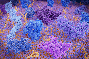
Advance Your Autoimmune Disease-Targeted Therapies
Our experts explain how combining disease-relevant models, in vitro inflammation models, and autoimmune T cell assays creates a clear picture of how your therapy is reprogramming the immune system.
Watch Now
As we see an increasing number of antibody-based therapies heading through the drug development pipeline, it is important not only to consider cross-reactivity onto mouse or proving efficacy in a mouse equivalent of your therapeutic, but also to test using the relevant human immune cell populations.
Our team has developed numerous pharmacology, efficacy, and mechanistical actions (MOA) immunology assays and animal models of inflammation and autoimmunity to support your drug development.
Understanding MOA Drives Model Selection
Charles River’s immunology platform combines autoimmune disease models, autoinflammatory pharmacodynamic (PD) and efficacy models with expertise in ex vivo analysis of immune cell subsets and primary human immune cell assays. In addition to guiding your model selection, our team can help you determine if you are engaging your target with the expected outcomes by evaluating the right ex vivo readouts and running relevant assays with human immune cell subsets.
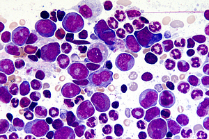
In Vivo Pharmacodynamic Models for Compound Selection
Pharmacodynamics models can drive your immunology assay, before entering lengthy and expensive disease-specific models. Examine the role of a therapeutic outside of the disease setting, to determine if it is hitting its target and modulating the desired cell type.
Download the Poster
Inflammation Model By Therapeutic Area
- Systemic In Vivo Models
-
Pharmacodynamic (PD) In Vivo Models
- DTH models
- T cell and B cell activation studies
- Inflammatory cell recruitment models
- LPS-induced cytokine release models
- NLRP3 inflammasome activation model
- Passive cutaneous anaphylaxis model
- Acute itch models
- Acute pain
- Acute inflammatory models
- Taste model
Learn more about Pharmacodynamic Models for Target Engagement
-
Gut
- In vivo models
- In vitro assays
- T cell assays
- Cytokine analysis
- Macrophage assays
- Neutrophil assays
- Epithelial assays
Learn more about IBD Mouse Models
-
Dermal
- In vivo models
- Atopic dermatitis models
- OXA-indued AD
- VD-induced AD
- OVA-induced AD
- HDM-induced AD
- Bleomycin-induced models of scleroderma
- Contact dermatitis models
- Imiquimod-induced psoriasis model
- Atopic dermatitis models
- In vitro assays
- T cell assays
- Cytokine analysis
- Macrophage assays
- Dendritic cell assays
- Neutrophil assays
- Innate lymphoid cells
- Epithelial assays
- In vivo models
-
Bone/Joint/Muscle
- In vivo models
- Collagen-induced arthritis models (mouse and rat)
- Collagen antibody-induced arthritis (CAIA) model
- CpG joint arthritis models
- CFA-induced inflammation
- Gout
- Carrageenan-induced inflammation
- Mono-iodoacetate joint osteoarthritis (OA) model
- ACLT (surgically induced OA model)
- DMM (surgically induced OA model)
- TPTX
- OVX/ORX
- In vitro assays
- Rheumatoid arthritis synovial fibroblast assay
- Macrophage assays
- B cell assays
- T cell assays
- In vivo models
-
Liver In Vivo Models
- CON A model
-
Kidney
- In vivo models
- Systemic lupus erythematosus (SLE) model
- Acute kidney injury - adriamycin induced
- Acute kidney injury - warm ischemia model
- Chronic kidney injury 5/6 and 3/4 nephrectomy
- In vitro assays
- Auto-antibody assays
- T cell activation assays
- B cell activation assays
- BUN and creatine analysis (ex vivo)
- In vivo models
-
Pancreas
- In vivo models
- In vitro assays
-
Lung
- In vivo models
- OVA pulmonary inflammation model
- LPS pulmonary inflammation model (ARDS model)
- Asthma models (ovalbumin, house dust mite, alternaria alternata)
- Bacterial lung infection models
- Lung fibrosis models (bleomycin)
- Poly:IC pulmonary inflammation model
- In vitro assays
- Cytokine analysis
- Macrophage assays
- Dendritic cell assays
- Mast cell assays
- Lung epithelial cell assays
- Endothelial cell assays
- B cell assays
- Basophil assays
- Neutrophil assays
- Eosinophil assays
- Fibroblast assays
Learn more about Respiratory Disease Pharmacology Studies
- In vivo models
-
Ocular
- In vivo models
- In vitro assays
- T cell proliferation/activation (ex vivo)
- Cytokine analysis
- Macrophage assays
Learn more about Ocular Disease Pharmacology Studies
-
Neuroimmunology
- In vivo models
- LPS model of microglial activation
- Experimental autoimmune encephalomyelitis (EAE) models (multiple sclerosis)
- In vitro assays
- Adult murine microglial assays
- Murine microglial: neuron co-cultures
- Human iPSC-derived microglia
Learn more about Neuroimmunology Studies
- In vivo models
-
Tumor
Learn more about Oncology & Immuno-Oncology Studies
-
Microbiome
The impact of therapies modulating the microbiome can be assessed across all autoimmune and inflammation models where the microbiome is thought to modulate disease progression and severity. We can support translation of your microbiome-directed therapy from culture of anaerobic bacteria, in vitro bacterial and viral assays and autoimmune/inflammatory/tumor efficacy models (e.g., diseases that impact the barrier epithelial layers, such as IBD in the gut or psoriasis of the skin).
In addition, in vivo infection models allow you to assess if your therapy modulates disease driven by pathogenic bacteria. 16S rRNA Sequencing can determine the impact of your therapy on microbiome diversity. Ex vivo readouts of immune cell function and histology support identification of whether the immune system has been modulated in the expected manner. In vivo model data can be supported by human epithelial, immune cell, bacterial co-culture assays to help translate to human cell biology and determine MOA.
Immunology assays have become an important tool for many types of drug development, including therapeutic areas not known to have an immune component. Charles River has developed extensive experience with those in vitro and in vivo immunology models and is able to advise on the best assays to collect human translational data based on the characteristics of your drug.
Frequently Asked Questions (FAQs) about Immunology Assays & Autoimmune Disease Models
-
Why target the immune system during drug development?
When conducting immunology research, it is critical to understand how the complex immune system affects the progression and treatment of disease.
-
How should I choose my autoinflammatory/autoimmune disease model?
The model you select should be dictated by the proposed mode of action of your therapeutic and immune cell type you are looking to target. IBD models, for example, are chemically induced models (e.g., DSS colitis) strongly driven by the innate immune system. Therefore, it would not be useful if your therapy was aimed at T cell regulation, in which case the adoptive transfer model of colitis would be more informative. In addition, if your therapy is aimed at driving epithelial repair, it will be necessary to choose a model that is able to resolve. In this case, the DSS colitis model may be a good choice, as upon withdrawal of DSS the epithelial barrier begins to repair, and several cycles of damage can be induced by reintroduction of DSS into the drinking water.
-
Are you able to support ex vivo readouts from your autoimmune disease models?
Yes, our immunology research team can support ex vivo readouts of immune cell profiling and function from a variety of tissues. This includes up to 18-color flow cytometry, analysis of cytokines, T cell proliferation in response of re-stimulation with the relevant peptide, and immune cell functional assays. In addition, we provide histological analysis.
-
How can in vitro human primary immune cell assays support my immunology research?
In vivo efficacy models demonstrate that you can inhibit disease progression and severity. However, the human immune system does differ from that of the mouse. Using primary human immune cell in vitro assays gives you data that allows you to translate the mode of action of your therapy into man. Assays can be simplistic in setup, such as polyclonal T cell assays, to more sophisticated multicellular routine or custom-built assay systems.
-
Why use pharmacodynamic (PD) models?
PD models are often an informative and quick way of screening your therapy to determine if it is on target in vivo. Often driven by a specific cell type, these assays can help narrow down lead candidate choices during your immunology research before moving to more lengthy, complex, and costly efficacy models.
-
How can you help progress my microbiome therapy?
Our team of immunologists and microbiologists works together in a multidisciplinary approach to support your microbiome therapy development. Our experience across infection, tumor, autoimmune disease, and inflammation models both in vivo, ex vivo, and in vitro allows us to support your immunology research program across multiple stages and readouts.