
Why Choose the 5xFAD Mouse Model for Alzheimer’s Research?
The 5xFAD mouse model of Alzheimer’s disease is a transgenic model that overexpresses two key human proteins with mutations related to familial Alzheimer’s disease (FAD); amyloid precursor protein (APP) with Swedish (K670N/M671L), Florida (I716V), and London (V717I) mutations, and presenilin 1 (PSEN1) with M146L and L286V mutations. Expression of both transgenes in 5xFAD mice is regulated by a Thy1 promotor to drive overexpression in neurons. 5xFAD mice recapitulate many aspects of cognitive and pathological changes observed in AD.
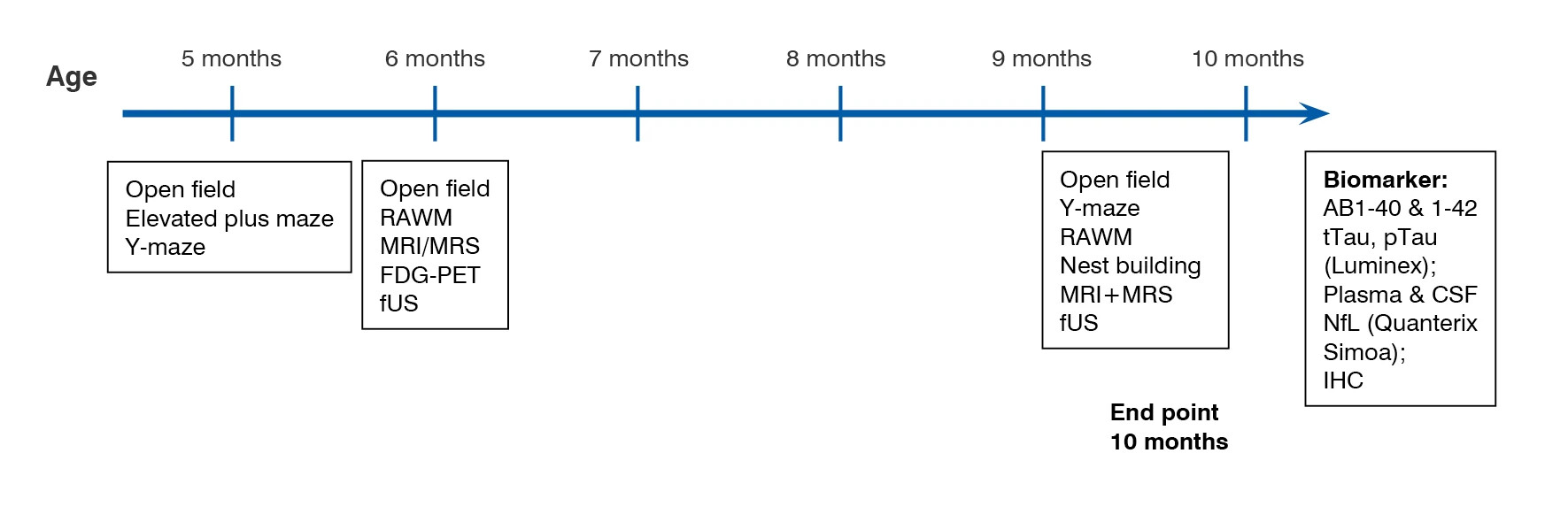
At Charles River, the 5xFAD mouse model has been validated using multiple endpoints including cognitive and behavioural testing, in vivo imaging, and analysis of biomarkers, such as amyloid plaque load, insoluble and soluble amyloid species, neurofilament light chain levels, and markers of neuroinflammation. Our validation data indicates that the 5xFAD mouse model displays significant cognitive deficits, reduced whole brain volume, CNS metabolism and neurometabolite profile changes, as well as age-dependent amyloid pathology.
-
Behavioural Testing
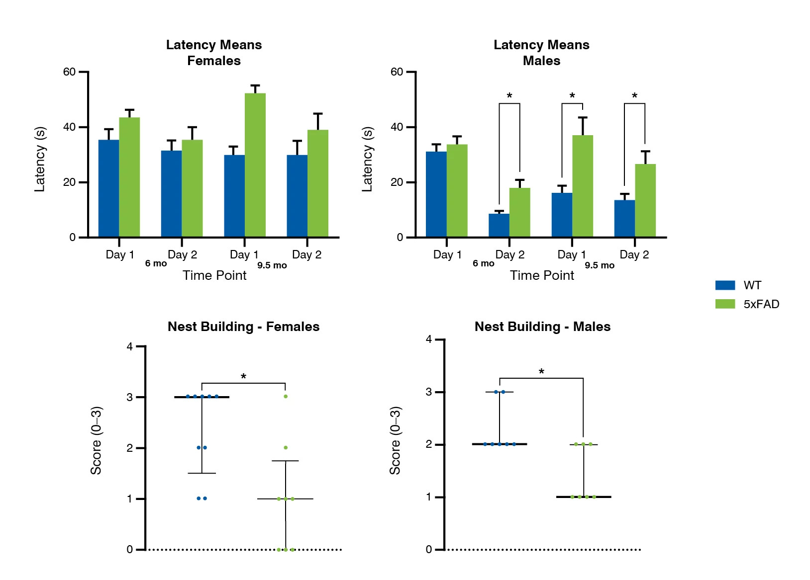
Male 5xFAD mice exhibited significant cognitive deficits in the radial arm water maze, used to measure spatial learning and memory, at 6 and 9.5 months of age. Additionally, both male and female 5xFAD mice showed impaired nest building performance, which is suggested to be sensitive to hippocampal function. In other behavioural tests used 5xFAD mice showed no significant phenotype; this included open field testing, elevated plus maze, and Y-maze.
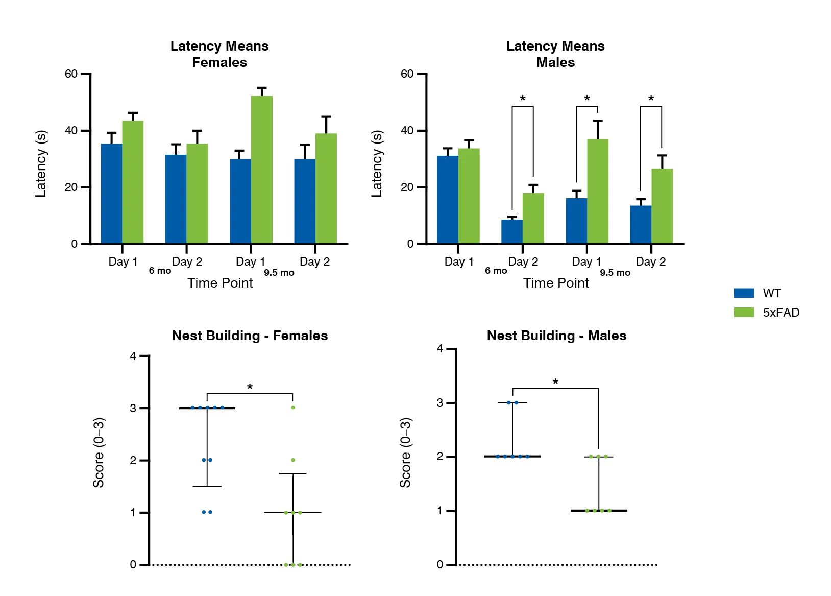
Figure 1: Cognitive deficits in 5xFAD Alzheimer’s disease mouse model, measured by radial arm water maze, and nest building.
-
In Vivo Imaging
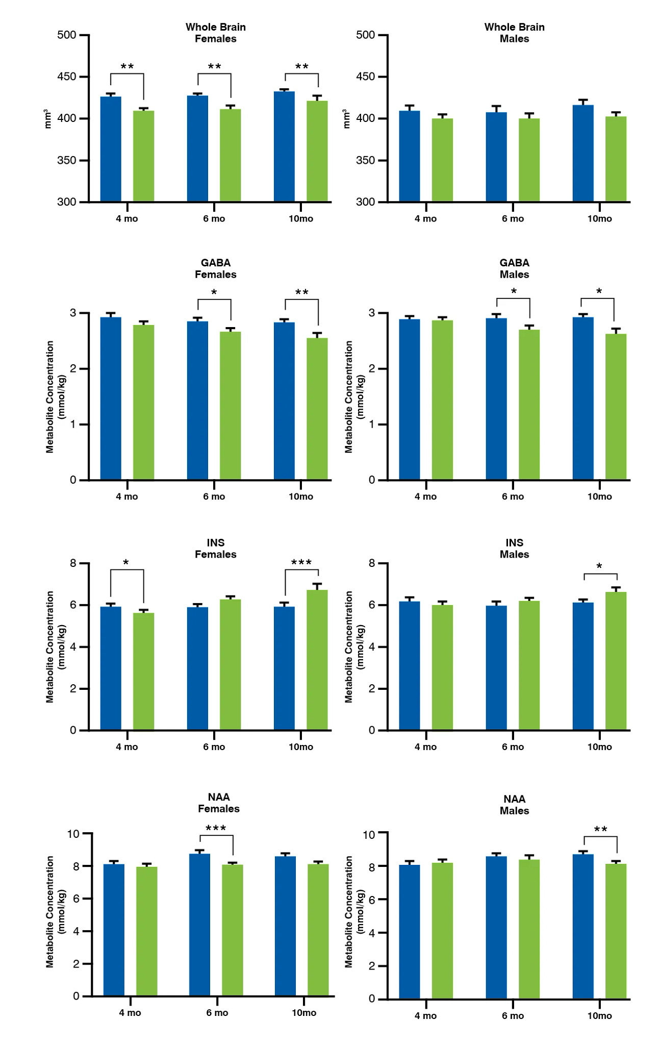
Various imaging techniques were used to investigate the 5xFAD mouse model. Whole brain volume, determined by magnetic resonance imaging (MRI) showed significantly reduced brain volume in female 5xFAD mice at all ages tested (4, 6, and 10 months).
Magnetic resonance spectroscopy (MRS), used to qualitatively evaluate metabolic markers, showed that the hippocampal neurometabolite profile (GABA, myo-inositol, and NAA) of 5xFAD mice was altered from 6 months of age. The increase of myo-inositol/NAA ratio observed in 5xFAD mice also serves as clinical biomarker for progression of mild cognitive impairment (MCI) to Alzheimer’s disease and of severity of Alzheimer’s disease.
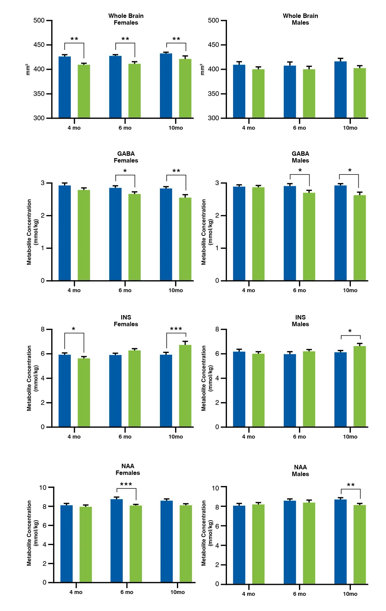
Figure 2: Brain volume and metabolite profile in 5xFAD Alzheimer’s disease mouse model, measured by MRI and MRS, respectively.
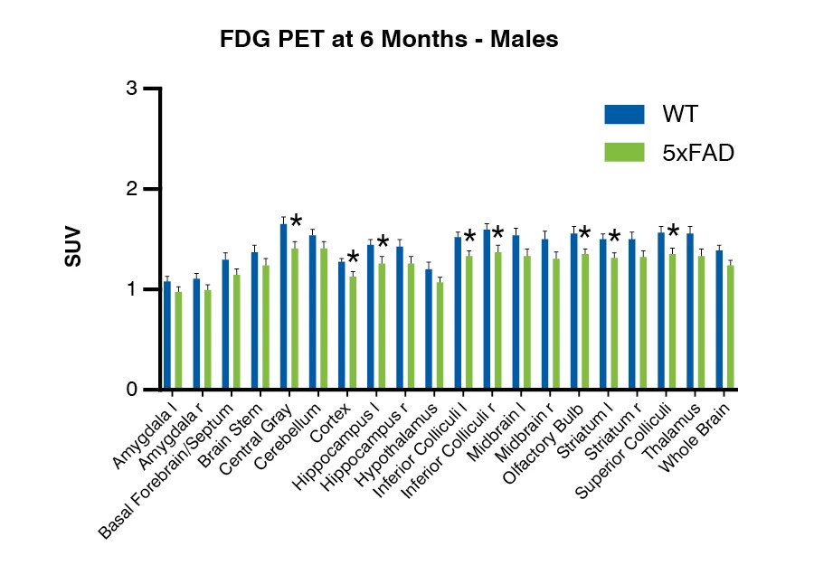
Additionally, PET imaging using 18F-fluorodeoxyglucose (FDG) to measure tissue metabolism showed that 18F-FDG uptake was significantly reduced in several brain regions from 6 months of age, suggestive of impaired brain glucose metabolism.
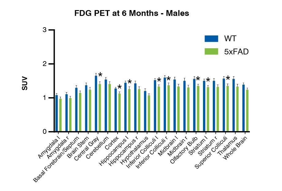
Figure 3: Brain metabolism in 5xFAD Alzheimer’s disease mouse model, measured by FDG PET imaging.
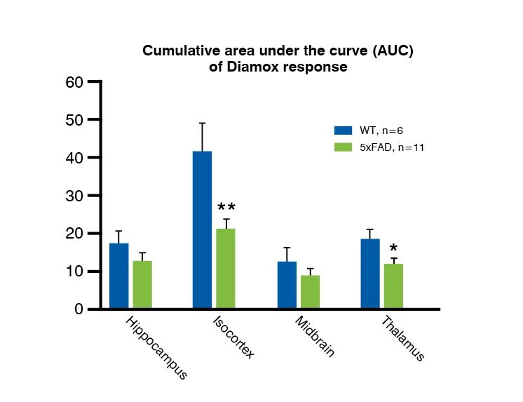
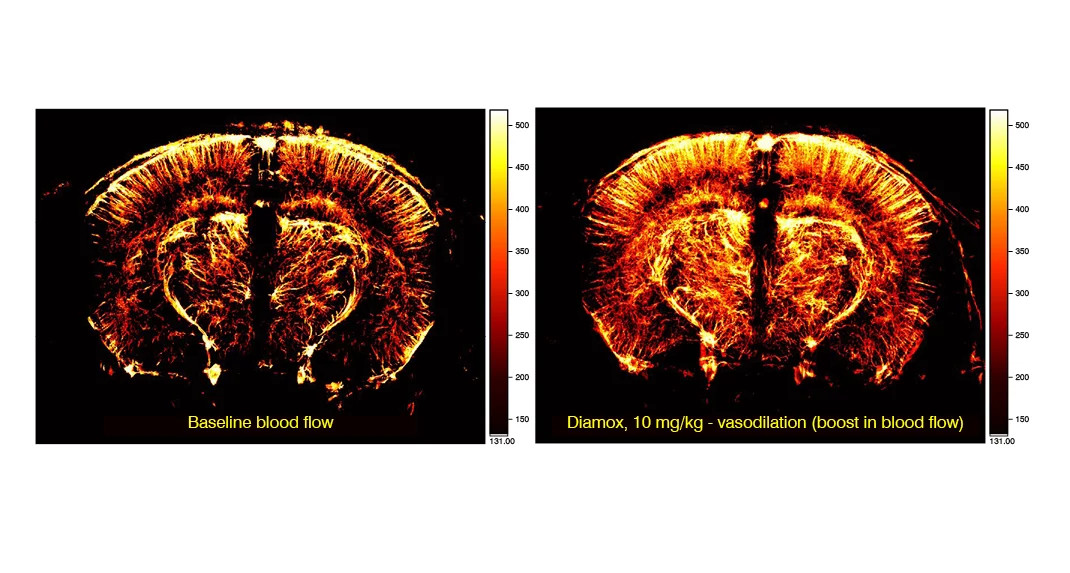
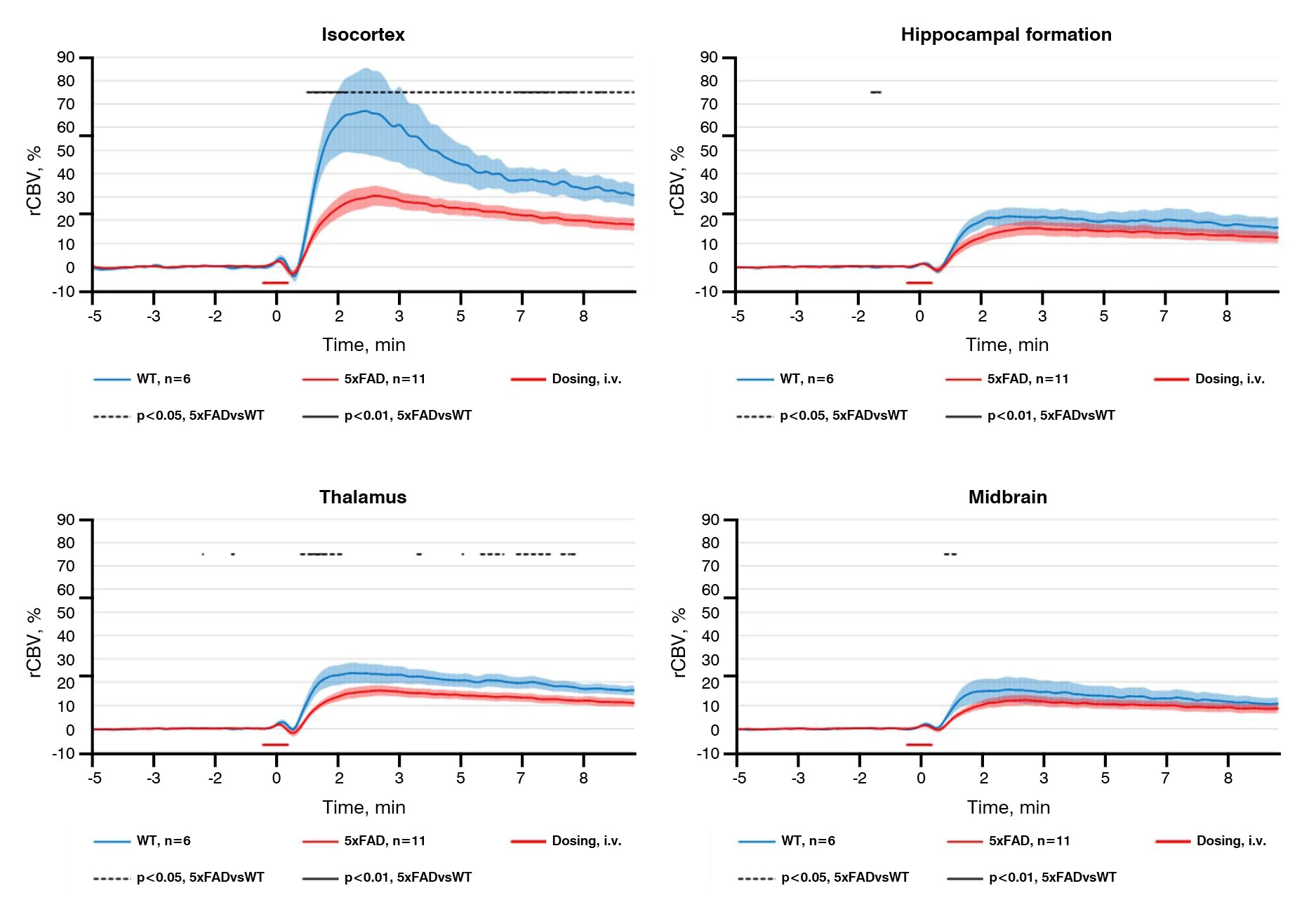
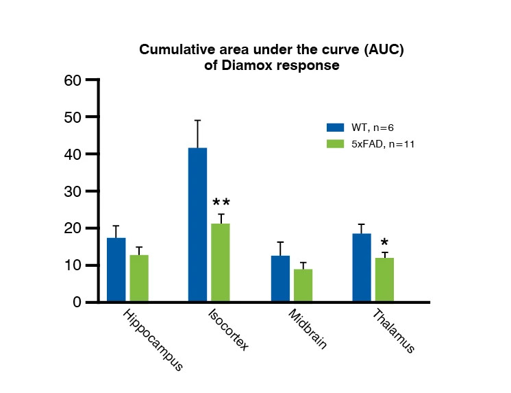
Functional ultrasound imaging, used to investigate blood volume changes and vascular reactivity, shows that 5xFAD mice have deficits in vascular reactivity, shown by impaired response to Diamox treatment, in several brain areas from 10 months of age. These results are in line with observations of reduced reactivity due to vascular amyloid pathology in other Alzheimer's disease mouse models and in patients.
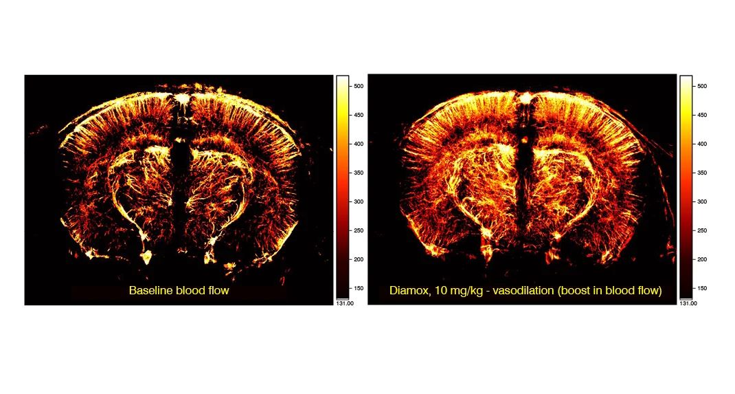
Figure 4: Median group map of the cerebral blood flow in 5xFAD Alzheimer’s disease mouse model, derived from a single microbubble tracking. Treatment with Diamox causes significant vasodilation, which is analysed to determine vascular reactivity.
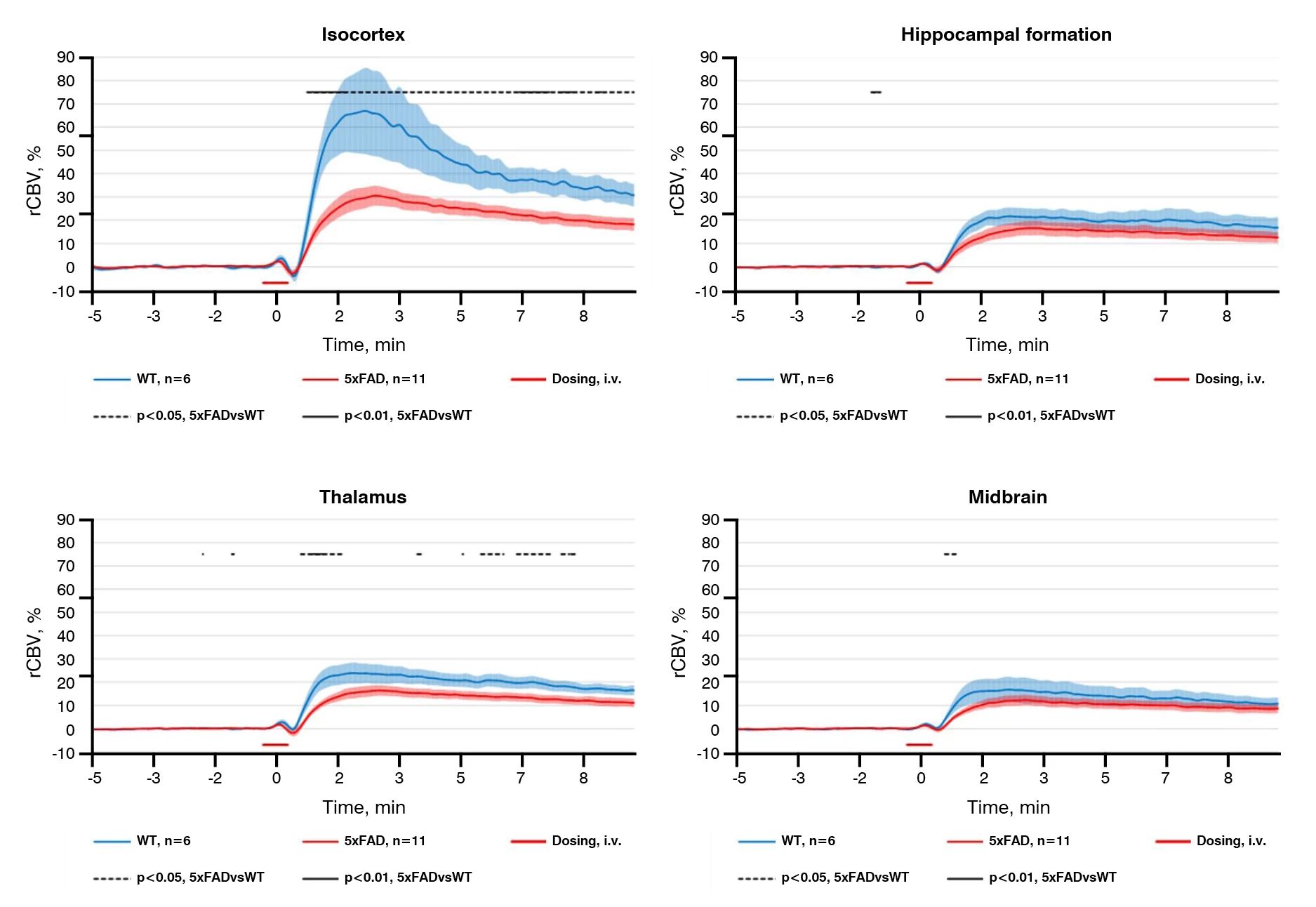
Figure 5: Vascular reactivity in 5xFAD Alzheimer’s disease mouse model, measured by functional ultrasound.
-
Biomarker Analysis
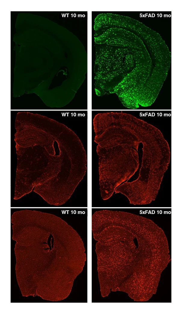
Various assays used to assess biomarkers in the 5xFAD mouse model of Alzheimer’s disease demonstrate that these mice develop strong, age dependent plaque formation with associated neuroinflammation and neurodegeneration:
- Aβ40 (soluble and insoluble) and Aβ42 (soluble and insoluble), determined by Luminex assay, were significantly increased at 6 and 10 months of age, in the hippocampus and parietal cortex
- Immunohistochemistry showed significantly increased amyloid plaque pathology in the cortex, hippocampus and whole brain of 5xFAD mice
- Neurofilament light chain (NfL) was significantly increased at 6 and 10 months of age, in both plasma and CSF
- Immunohistochemistry markers showed significant astrogliosis and microgliosis in the cortex, hippocampus and whole brain of 5xFAD mice
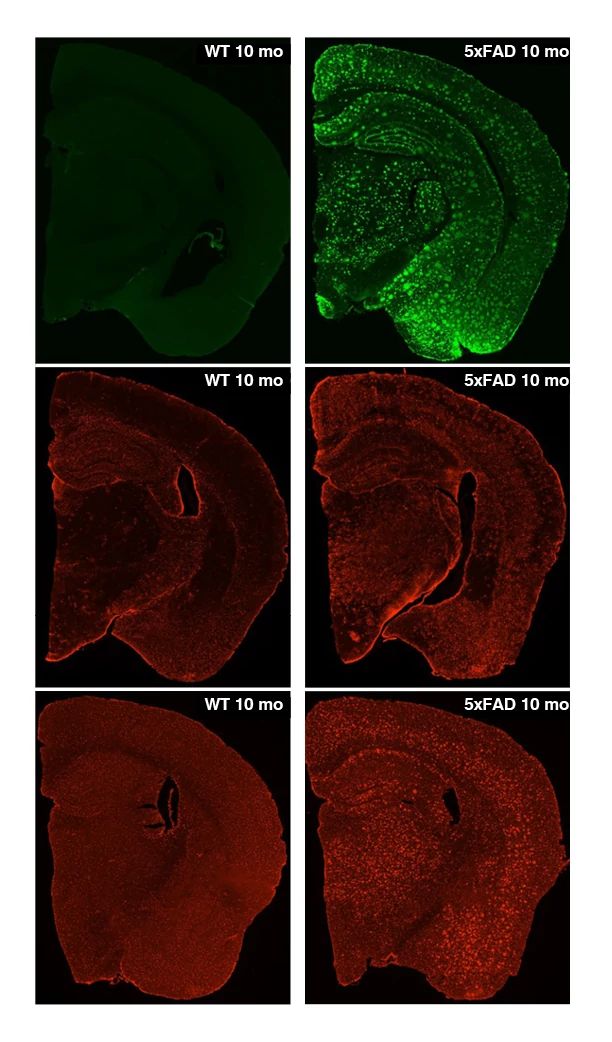
Figure 6: Biomarker analysis in 5xFAD Alzheimer’s disease mouse model, showing increased amyloid β deposition (top panels), GFAP staining for astrocytes (middle panels), and Iba1 staining for microglia (bottom panels).
Please note, the 5xFAD and Tg2576 mouse models are only available for use in studies conducted through Charles River Labs; they are not available to purchase independently.
Exploratory Toxicology for Neuroscience Drug Discovery
This eBook describes strategies across the early stages of drug discovery to support confidence in your lead small molecule candidate and to ensure you proceed through the drug development process with the most promising candidate.
Download eBook
Components for Your 5xFAD Mouse Model Alzheimer’s Disease Studies
Behavioral Testing
- Open field test
- Nest building
- Contextual fear conditioning
- Elevated plus maze
- Y maze
- Radial arm water maze
In Vivo Imaging
- MRI for whole brain volume
- Proton magnetic resonance spectroscopy (MRS) for metabolic profiling
- Fluorodeoxyglucose (FDG) PET imaging for brain metabolism
- PET imaging for neuroinflammatory markers
- PET imaging for GPCR activation or receptor occupancy
- Functional Ultrasound imaging for vascular reactivity
Biomarker Analysis
- Soluble and insoluble Ab40 and Ab42 in brain tissues, plasma and CSF
- Neurofilament light (NfL) in brain tissues, plasma and CSF
- Plaque load in brain tissues by immunohistochemistry
- Astrogliosis and microgliosis by immunohistochemistry
- Cytokine profiling by ELISA
- Neurotransmitter and neurochemical profiling by microdialysis (ACh, 5-HT, NE, DA, Glu, metabolites)
- Microdialysis for pharmacokinetic studies