
Preclinical Functional Ultrasound Imaging of the Brain
In the brain, functional ultrasound imaging measures transient changes in blood volume at better spatiotemporal resolution than any other functional brain imaging technique. The readout is based on the well-known doppler effect where waves are reflected from red blood cells moving through blood vessels. The signals are directly proportional to relative cerebral blood volume (rCBV). It is a novel technique that was developed to identify regions of brain activation. One of the first study’s to use fUS explored whisker-evoked cortical and thalamic responses of epileptiform seizures in the brain of an awake rat.
Figure 1: Structural fUS scan of a rat brain, 3D MIP

Poster: Functional Ultrasound Assessment of Seizure Threshold Modulation
Chronic epileptic animals have increased susceptibility to seizures. In this study, we used functional ultrasound (fUS), to record the brain hemodynamic response to a sub-convulsive dose of kainic acid (KA) - an epileptogenic chemoconvulsant.
View the Poster
Three Major Preclinical Functional Ultrasound Imaging Applications
There are three major ways functional ultrasound imaging can be used to characterize animal models of neurological and neurodegenerative diseases:
- Pharmaco-fUS: response to acute pharmacological challenge (e.g., nicotine cortical activation in mice) or altered receptor binding
- Stimulation-fUS: altered stimulus-evoked activation by loss and/or restoration of functionality (e.g., somatosensory whisker stimulation with electrical stimulation to study pathologies in the trigeminal pathway such as migraine and neuropathic pain)
- Resting-state-fUS: altered neurotransmitter systems and changes in functional connectivity (e.g., requires no stimulation, a relaxed animal to obtain resting state data with no interference from the environment)
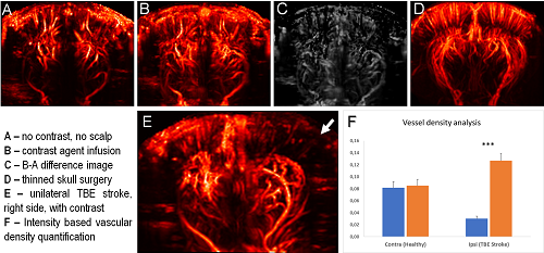
Figure 2: fUS is used as a structural imaging tool in rats for visualization of the ischemic area in thromboembolic stroke (TBE).
Frequently Asked Questions (FAQs) About Functional Ultrasound Imaging
-
What are the preclinical imaging advantages exist for that functional ultrasound imaging vs fMRI?
fUS imaging has five times the sampling rate and three times the spatial resolution compared to functional MRI (fMRI).
-
What are Charles River’s preclinical imaging capabilities around fUS?
Functional ultrasound imaging capabilities are fully developed at Charles River. A few pilot studies are underway such as the oxaliplatin-induced peripheral neuropathy model and thromboembolic stroke model. Other routine tests include somatosensory and electrical stimulations.
-
How long is the duration of protocol?
Charles River’s standard procedure takes approximately two hours to collect all three types of imaging data (pharmacological, somatosensory/electrical, and resting state).