
In Vitro Neuroscience Assay and Tom20 Staining
Parkinson’s disease (PD) pathogenesis has been linked to mitochondrial dysfunction through several lines of research, starting with the finding that the mitochondrial complex I inhibitor rotenone induces parkinsonism. In addition, mutations in genes encoding proteins involved in the selective clearance of dysfunctional and redundant mitochondria (mitophagy), such as PARK2 and PINK1, are present in most autosomal recessive cases of PD. Likewise, a growing body of evidence implicated mitochondrial dysfunction in other neurodegenerative disorders such as Alzheimer’s disease (AD), amyotrophic lateral sclerosis (ALS), and Huntington’s disease (HD). For instance, genetic perturbations in genes involved in mitophagy have been demonstrated to underlie ALS, whereas amyloid beta (Aβ) and hyperphosphorylated Tau-induced defective mitophagy has been shown to contribute to AD pathology. Phenotypic readouts to measure mitochondrial (dys) function in disease-relevant cellular backgrounds are therefore thought to represent powerful predictive tools to probe neurodegenerative pathobiology and identify potential therapeutics that can augment mitophagy.
TOM20 is a subunit of the mitochondrial translocase of the outer membrane (TOM) complex and represents a biomarker for mitochondrial abundance. By profiling therapeutic candidates in absence (mono-treatment) and presence (co-treatment) of an established mitophagy-inducing trigger, candidate molecules that enhance trigger-induced mitochondrial clearance without damaging mitochondria directly may be selected.
The Mitophagy TOM20 loss assay represents a scalable and fast neuroscience in vitro assay to potentially screen large compound sets for their ability to augment mitophagy in a neuronal background. The assay combines data-rich high content analyses with high-throughput image acquisition. This assay, and a complementary α-synuclein assay, are intended to evaluate small sets of potential lead series prior to larger lead-optimization endeavors and in vivo translation.
Mitophagy Assay Principle
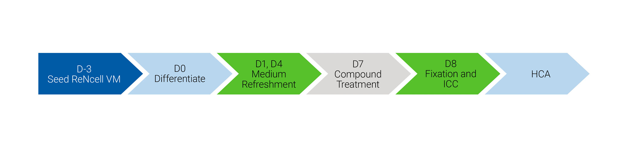
Immortalized human mesencephalic progenitor cells (ReNcell VM) are differentiated by withdrawal of growth factors (bFGF, EGF) and addition of pro-differentiation factors (cAMP, GDNF) for seven days. Cells are then treated with test compounds in the absence and presence of 1 μM oligomycin/antimycin (O/A; a commonly used mitophagic trigger) for 18 hours followed by immunocytochemical TOM20 staining. Combining treatment of mitophagy-enhancing compounds with 1 μM O/A leads to a dramatic reduction in TOM20 levels on top of reductions induced by O/A treatment alone. TOM20 immunostaining intensity is quantified using in-house developed high content analysis-based (HCA) algorithms. Nuclear counts are quantified to identify potential compound-induced cytotoxicity.
TOM20 Assay Setup
- Cell type → ReNcell VM cells
- Seeding density → 50,000 cells/well in laminin-coated 96-well plates
- Differentiation → Differentiation medium (-bFGF/EGF, +cAMP/GDNF) on D0, with refreshments on D1 and D4
- Compound Treatment → 8-point CRC on D7, both in absence and presence of 1 μM O/A
- Assay controls → 1 μM O/A (positive control) and .1% DSMO (negative control) on D7
- Readout → HCA-based quantification of TOM20 immunoreactivity
- Immunocytochemistry → Fixation after 18h treatment, anti-TOM20, and DAPI staining on D8

Explore AD/PD 2025
The AD/PD™ Conference is where the brightest minds gather to explore the latest research, clinical trials, and therapies for Alzheimer's and Parkinson's diseases, from uncovering groundbreaking discoveries to sharing the latest developments in treatments. You are sure to find new insights to help you through your discovery and development process.
Browse Resources
Mitophagy Assay Performance
Representative data for the Mitophagy TOM20 loss assay is shown below.
Immunocytochemical Staining of Differentiated ReNcell
VM Cells Using an Antibody Directed Against Human Tom20
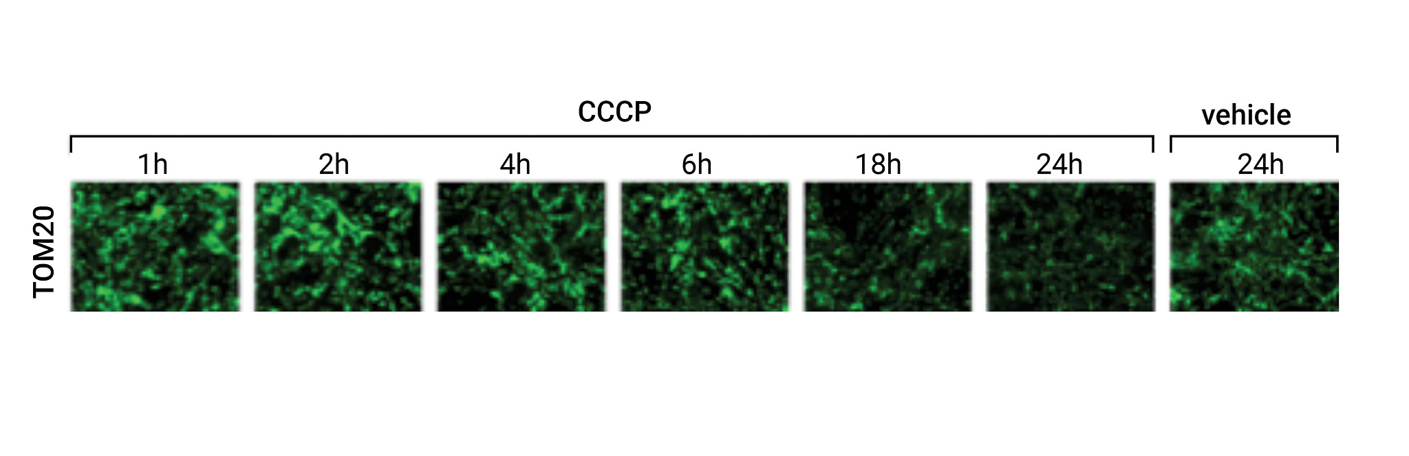
The mitochondrial uncoupler CCCP (10 μM) or vehicle was added for the indicated treatment times followed by immunocytochemical analysis using mouse anti-TOM20.
Assay Validation Using Prototypical Mitophagy Triggers
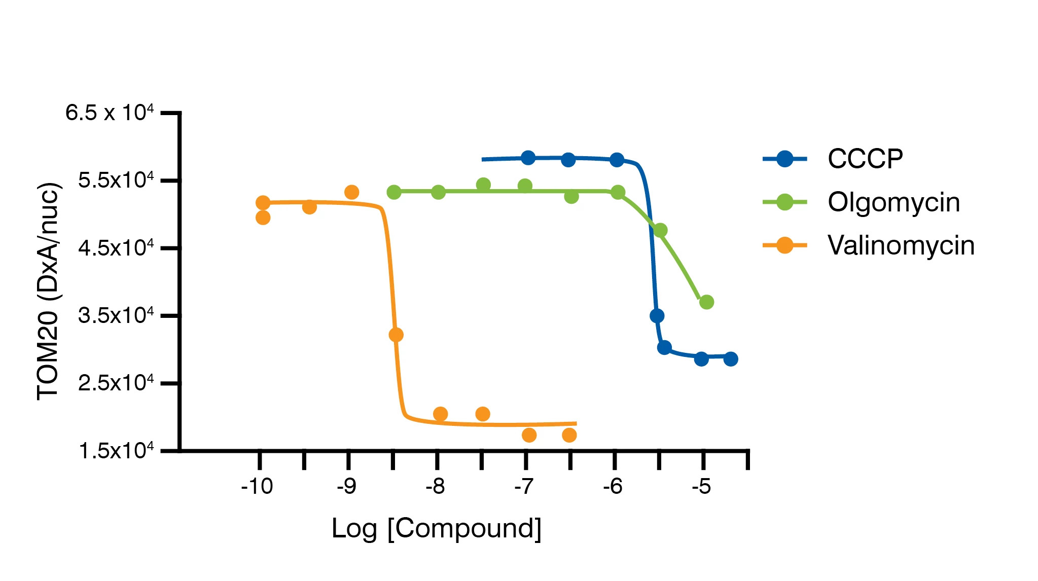
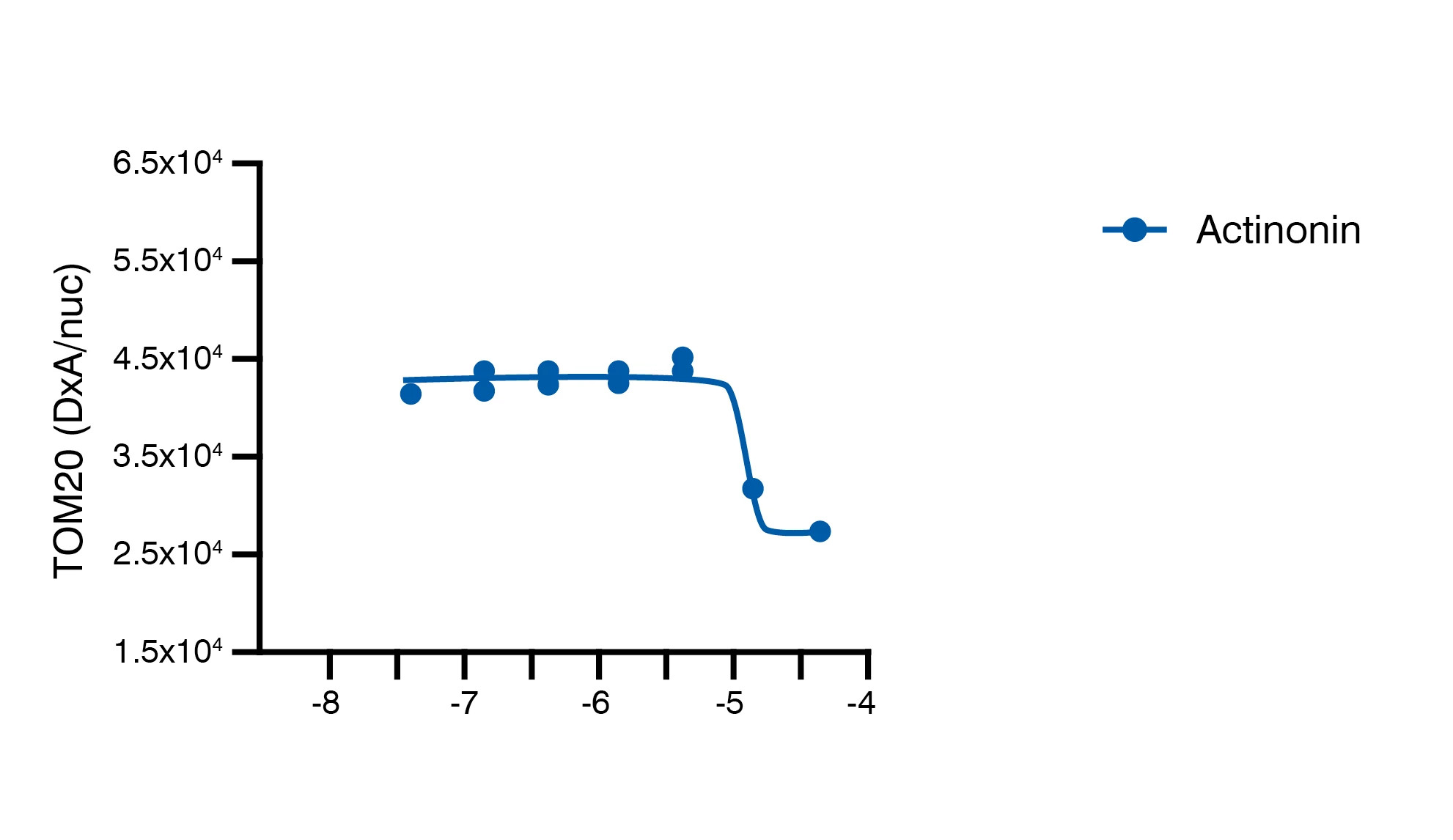
Differentiated ReNcell VM cells were treated with increasing concentrations of CCCP, oligomycin, valinomycin or actinonin for 18 hours followed by immunocytochemistry using mouse anti-TOM20 antibody and HCA-based quantification of TOM20 immunoreactivity.
Summary of Tom20 Mitophagy Assay
The therapeutic candidates are evaluated in 8-point concentration response curves at 0.5Log-fold dilution steps in biological triplicates in the absence and presence of 1 μM O/A. In addition, potential cytotoxicity of the tested therapeutic candidates is assessed by HCA-based quantification of nuclei loss derived from DAPI-staining. As a positive control for Mitophagy TOM20 loss, 1 μM O/A is tested on each plate as a benchmark for assay quality. Results are provided as percentage effect (PE) and percentage remaining cells
Complementary Neuroscience Assays to Tom20 Mitophagy Assay
Alpha-Synuclein Expression and Aggregation Quantification