
T Cell-Mediated Chemotaxis
Chemotaxis assays offer the opportunity to evaluate drug candidates with possible chemoattractant properties that can impact on the migration of immune cells and in turn influence their ability to kill cancer cells. Chemotaxis is the directed movement of cells toward a chemical gradient. This plays an important role in biological processes such as tumor metastasis and immune responses. Chemokines are a group of structurally related cytokines that induce directed migration of various populations. Chemokine receptors induce cell movement toward a concentration gradient of the associated chemokine ligand. Chemokines are essential for the migration and tissue localization of various lymphocyte subpopulations expressing specific chemokine receptors. Human stromal cell-derived factor-1 SDF-1(α), also known as CXCL12, binds and signals solely though chemokine receptor CXCR4.
T Cell-Mediated Chemotaxis Assay Principle
Chemotaxis assays are conducted using IncuCyte® Clearview 96-Well Cell Migration Plates. The membrane of the upper insert is coated on the insert side and reservoir side with a fibronectin extracellular matrix. Freshly isolated human PBMCs from healthy donors or Jurkat cells are seeded (80 µL/well, 5000 cells/well) into the IncuCyte® Clearview insert. If the assay requires additional treatment, the seeding volume is reduced to 60 µL. A 4x concentration of treatment is prepared, and 20 µL added to the insert wells and pre-incubated with the cells prior to exposure to chemoattractant. Cells are allowed to settle for 45 to 60 minutes on a level surface at ambient temperature. The insert plate containing cells is placed into the reservoir plate filled with 200 µL of chemoattractant or controls. The plate is imaged on the IncuCyte® at intervals using phase contrast to provide direct visualization of cell migration from the insert plate to the reservoir.
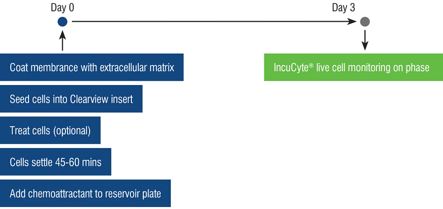
T Cell-Mediated Chemotaxis Assay Set Up
A chemotaxis protocol has been developed for optimum analysis of cell migration using PBMCs or Jurkat cells in response to a fibronectin extracellular matrix and the chemoattractant SDF-1(α).
| Chemotaxis Assay |
|---|
| Donor → PBMCs |
| Cell Line → Jurkat |
| Seeding Density → 5000 c/w |
| Coating → Fibronectin 5 µg/mL |
| Chemoattractant → SDF-1α (31.25 - 125 nM) |
| Negative Control → RPMI 0.5% FBS |
| Monitor → Scan every hour |
| Readout → Total phase object area normalized to initial top value |
Performance
Representative data is shown below for Jurkat cells and human PBMCs seeded onto a fibronectin (5 µg/mL) coated Clearview plate. Cell migration is monitored by movement of cells through the pores of the membrane on the upper insert to the lower reservoir containing chemoattractant. The data is generated and analyzed with the IncuCyte® S3 Live-Cell Analysis system which automatically acquires images over time. Data is expressed as total phase object area normalized to the initial top value.
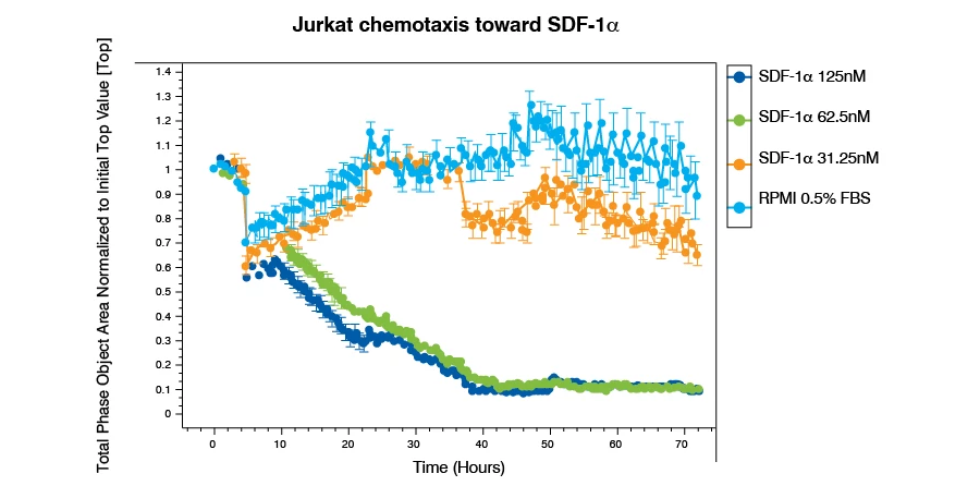
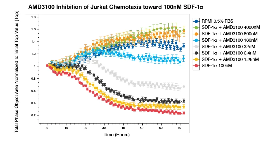
Jurkat cells demonstrate a concentration-dependent migration towards the CXCR4 agonist SDF-1α chemoattractant. The response with 100 nM SDF-1α can be inhibited with the selective CXCR4 inhibitor AMD3100.
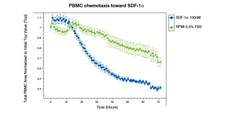
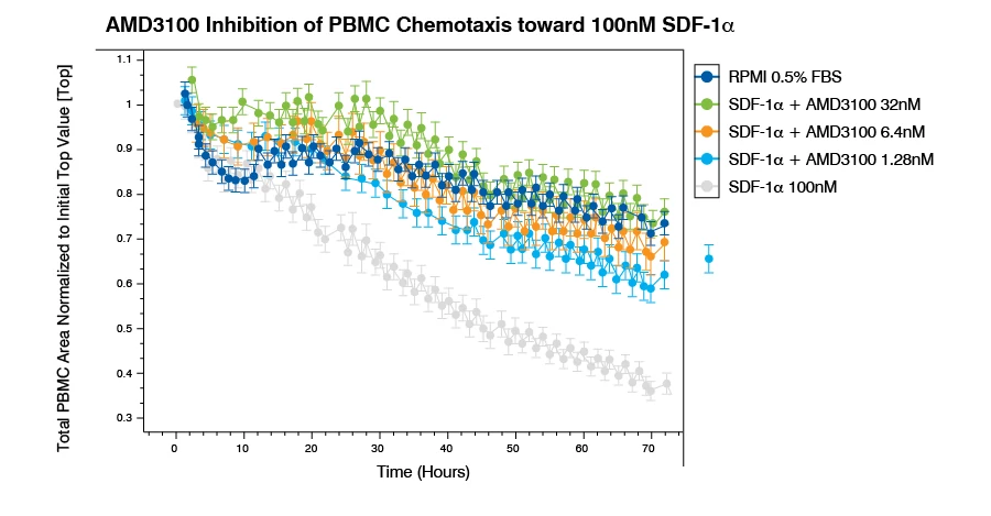
Human PBMCs demonstrate migration towards 100 nM SDF-1α chemoattractant. The response can be inhibited with the selective CXCR4 inhibitor AMD3100.
Summary
Here we show immune cell chemotaxis using the line of immortalized T lymphocyte Jurkat cells and isolated human PBMCs in response to α (CXCL12), the ligand for CXCR4. The loss of cell area from the top of the membrane indicates migration of the T cells towards the CXCL12 ligand by interaction with CXCR4 receptor on the cell surface. PBMCs demonstrate a slower rate of migration compared to the Jurkat cells.
Chemotaxis toward a maximal concentration of α is inhibited by the selective CXCR4 inhibitor AMD3100 in a concentration-dependent manner for both Jurkat cells and human PBMCs.
Using this assay, changes in cell migration due to immune enhancement or immune suppression by therapeutic antibodies, T cell therapies (CAR-T cells), and small molecules to target tumor cells may be identified using isolated primary human PBMCs.
Need a custom version of this assay?
Let us know how we can customize this in vitro assay to better suit your target validation.
How Can We Help