
Comprehensive Ocular Pathology Study Solutions
Embarking on an ocular pathology path requires delving into areas marked by complexities and specificities. It’s a field where specializations intersect, and where the need for genuine expertise and reliable solutions is critical. At Charles River, our mission extends beyond proficiency; we aim to deliver unapparelled, comprehensive solutions in ocular pathology studies, establishing a standard for precision and reliability across every segment of your research.
Our team of ocular drug development experts can provide personalized guidance and support on techniques and methodologies needed for your ocular pathology needs throughout your journey. We’re not just navigating this landscape, we’re helping redefine it, elevating standards of excellence.
With our state-of-the-art facilities and advanced technology platforms, we are well-equipped to support all your research needs throughout your ocular study. Whether your research requires the application of time-tested methodologies or guidance on rigorous pathological evaluation, we are here to help.

Webinar Series: Are You Ready for the Digital Pathology Revolution?
Digitalization is revolutionizing traditional laboratory pathology. Potentially more objective, accurate, and faster than traditional microscopy, this technology is changing our workflow.
Watch Now
Key Areas of Our Expertise in Ocular Pathology Studies
Expert Ocular Pathologists
Our team of ocular pathologists are not just specialized in general pathology but are uniquely focused on ocular toxicology. This specialization enables us to discern subtle changes and patterns specific to eye tissues, which is crucial for the accurate assessment of ocular toxicity studies. Their expertise is pivotal in diagnosing and understanding the progression of ocular diseases, leading to more informed decisions in the development of ocular treatments.
Advanced Ocular Histology
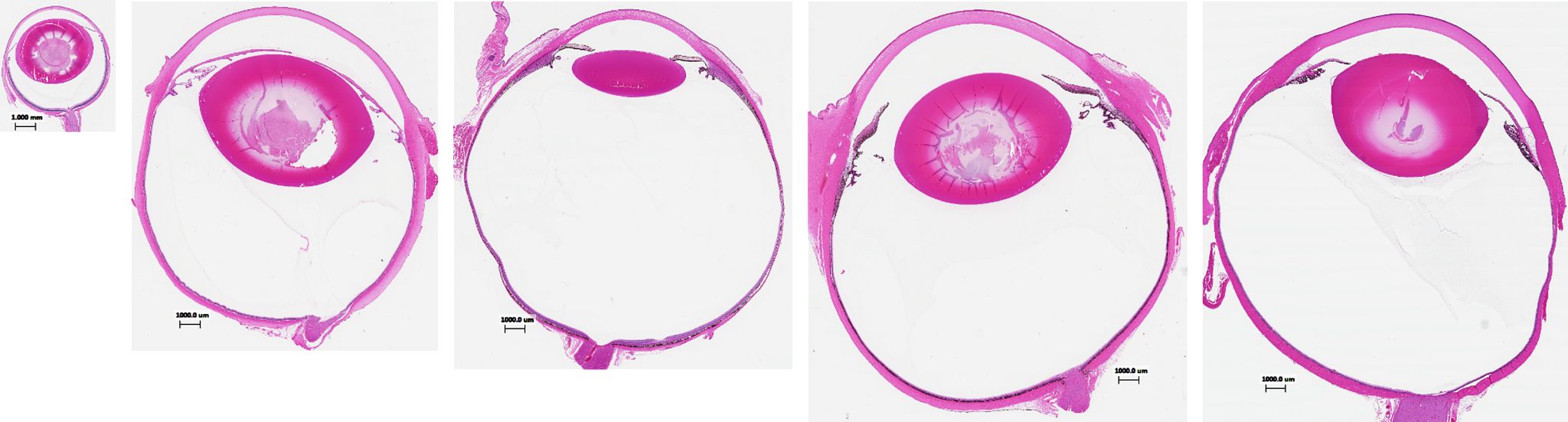
With years of accumulated experience, our lab has perfected techniques to prepare eye tissue samples that reveal the most intricate details under microscopic examination. This includes precise sectioning tailored to the specific needs of each study, ensuring that every relevant anatomical feature is captured with the utmost clarity. Our meticulous preparation of tissue samples sets the foundation for accurate histopathological evaluation, providing a detailed understanding of the ocular effects of your test item.
Molecular Pathology Techniques
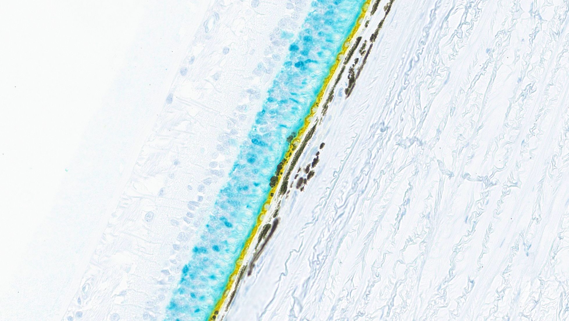
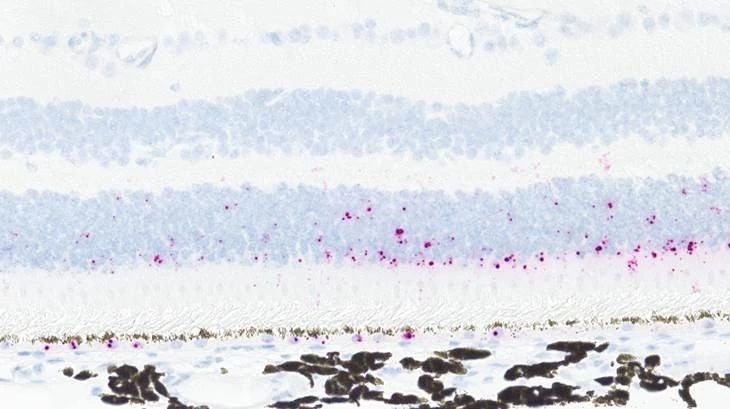
Our molecular pathology capabilities allow for the precise localization and identification of proteins and nucleic acids within the eye, which is essential for understanding the molecular underpinnings of ocular diseases and the effects of therapeutic agents such as gene and cell therapies. Techniques such as:
- Immunohistochemistry (IHC): We utilize targeted antibodies to stain specific proteins within eye tissues, providing insights into the pathophysiology of eye diseases and the efficacy of therapeutic agents.
- In-situ hybridization (ISH): Our ISH techniques enable the visualization of gene expression patterns within eye tissues, shedding light on genetic factors that may influence disease progression and treatment outcomes.
Microdissection for Bioanalysis Assays (Including PCR)
Our skilled necropsy technicians perform microdissection with the utmost precision to isolate individual ocular structures for bioanalysis. This meticulous process ensures that we can accurately quantify the presence and concentration of your test item in each distinct part of the eye, providing a comprehensive profile of its distribution and potential efficacy.
Digital Pathology
We developed a sophisticated deep learning algorithm designed to quantify retinal atrophy. This tool is invaluable in our rodent models of retinal damage, providing objective and precise measurements of retinal health. The algorithm aids in demonstrating the neuroprotective properties of your test item, offering a quantitative approach to evaluating potential treatments for retinal diseases.
Elevate Your Ocular Pathology Study
Charles River is known for its experience and agility in delivering a wide range of solutions. We understand the diverse needs and complexities of ocular studies, and we are committed to ensuring your research goals are met with meticulousness and the highest standards of excellence.
Our team of seasoned experts is dedicated to propelling your research to new heights, offering insights sourced from the extensive depth of our collective knowledge and expertise.
Collaborating with us will cultivate innovation and advanced knowledge in ocular pathology – pushing the boundaries on not only what is possible today but critical in shaping what is possible in the future. Partner with us today.
Frequently Asked Questions (FAQs) About Ocular Pathology Studies
-
How does ocular histology play a role in ocular pathology studies?
Ocular histology is integral to our services, allowing for the detailed study of the microscopic structure of eye tissues. It aids in providing deeper insights in the cellular and tissue-level responses in ocular studies, contributing to the advancement of ophthalmic research.
-
How can ocular pathology help in the advancement of ophthalmology?
Ocular pathology advances ophthalmology by providing in-depth analysis of eye tissues, enabling the detection, and understanding of various ocular diseases and conditions. It aids in the development of therapeutic strategies and novel drugs, ensuring their safety and efficiency for combating eye diseases and promoting ocular health.
-
What kind of ocular toxicity studies are performed in ophthalmology?
We conduct comprehensive ocular toxicity studies such as intravitreal injections, topical instillation, intravitreal implants and dosing, subretinal/suprachoroidal dosing, and systemic administration. These studies encompass a range of assessments like microscopic examination of ocular tissues, histopathological evaluation, and special stain assessments. These studies are vital in identifying potential adverse effects of drugs on ocular structures and are integral to ensuring the safety of new ophthalmological interventions.