
Proteomics-Enabling Spatial Pathology Services for Smarter Decision Making
Your translational research depends on understanding how biology plays out within the tissue microenvironment; yet generating reliable, high‑plex spatial data can be complex. Our integrated spatial pathology platform is built to simplify that process for you. Through advanced multiplex staining technologies, 5–60+ biomarker panels, whole‑slide or region-of-interest (ROI)‑guided imaging, centralized image management, and AI‑supported analytics, we help you move efficiently from raw images to actionable, decision-enabling insights. This end‑to‑end approach provides the spatial resolution and reproducibility you need to link tissue context with functional outcomes across discovery and safety programs.
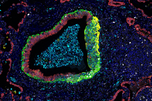
Advance Your Translational Research with High-Plex Spatial Profiling
This webinar will help you reveal deeper biological insights that accelerate decisions. Discover high‑plex spatial profiling and how it strengthens your translational research.
Watch Now
What You Gain from an Integrated High‑Plex Spatial Pathology Approach
High‑plex spatial pathology unlocks a deeper understanding of tissue biology by revealing what is present and where, through quantitative analysis of dozens to over 100 protein markers in their native spatial context. Using formalin-fixed paraffin-embedded (FFPE) and/or fresh‑frozen samples, we seamlessly integrate discovery, translational, and safety workflows with digital pathology, validated multiplex assays, and advanced biomarker analytics, enabling faster, more informed decisions.
Spatial proteomics extends genomics by mapping protein networks directly within tissue, revealing cell‑to‑cell interactions that genomics alone cannot capture. This added context strengthens biomarker discovery, mechanism‑of‑action (MoA) studies, and responder stratification. Our spatial profiling services bring these insights to translational programs where location and cellular proximity are critical to your research.
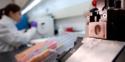
Sample Flexibility
FFPE, fresh‑frozen, and tissue microarrays (TMAs) align with standard histology workflows to preserve morphology and enable consistent analysis.
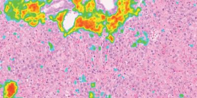
End‑to‑End Digital Pathology
Whole‑slide scanning, centralized image management, and AI‑assisted analytics integrate across our global pathology network to accelerate review and collaboration.

Translational Biomarker Expertise
Our deep experience in immuno‑oncology and beyond ties spatial insights to pharmacology, biomarkers, and study endpoints.
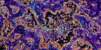
Spatial Proteomics Analysis Enablement
Enablement of comprehensive spatial proteomic analysis using leading technologies such as multiplex immunofluorescence (mIF) and digital spatial profiling (DSP).
An End‑to‑End Spatial Pathology Experience That Accelerates Insight
Our high-plex spatial pathology workflow doesn’t just give you data – it gives you clarity, confidence, and momentum. The result is a streamlined, end-to-end spatial profiling experience that elevates the impact of every study and every milestone.
Study Design and Panel Strategy
Define scientific questions, tissues, and ROI. We tailor panel depth (e.g., checkpoint, stromal, and functional markers) to study objectives and throughput needs.
High-Plex Acquisition
Execute multiplex immunofluorescence (mIF), including TSA-based, where appropriate, and digital spatial profiling (DSP) workflows with whole-slide or ROI-guided imaging. Commonly used platforms can support panels from five to 60+ platforms.
Analysis and Interpretation
Cell segmentation, phenotyping, neighborhood maps, and spatial statistics with audit-ready deliverables seamlessly integrate into discovery or safety assessment programs.

Your Essential Guide to Digital Pathology
Our Digital Pathology Services and Solutions interactive eGuide will help you understand the transformative power of our digital solutions to your pathology needs—because every moment matters.
Download your copy
How High‑Plex Spatial Profiling Advances Your Discovery and Safety Decisions
Our high‑plex spatial pathology services give you the context you need to accelerate discovery, deepen translational insight, and evaluate safety with greater precision. By pairing advanced spatial proteomics with robust digital analytics, we deliver the clarity and confidence to move your program forward, supported by comprehensive visualizations and analysis‑ready data.

Immuno-Oncology Biomarker Discovery and Responder Enrichment
Map immune contexture (e.g., PD1/PDL1 neighborhoods, Treg–CD8 proximity) to inform trial design.

MoA and Combination Therapy Profiling
Spatially resolve target engagement, pathway activation, and macrophage/T cell dynamics.

Safety and Translational Pathology
Integrate special stains, ISH, and high-content image analysis with GLP-aligned workflows where appropriate.

Data and Reporting
You’ll receive whole slide images, analysis-ready files, cell-level feature matrices, annotated phenotypes, spatial metrics, and publication-quality figures, with options for interactive review and secure sharing.
Frequently Asked Questions (FAQs) for High-Plex Spatial Pathology and Profiling Services
-
What is high-plex spatial pathology and why is it valuable?
High-plex spatial pathology maps dozens to over 60 protein markers in situ at single cell resolution, revealing cell types, functional states, and cell–cell interactions that drive disease and treatment response. These insights go beyond genomics alone for biomarker discovery and translational decision-making.
-
Which technologies underpin your multiplex immunofluorescence services and spatial profiling services, and how many markers/sample types are supported?
Barcoded cyclic IF platforms image over 60 proteins on whole slides; digital spatial profiling quantifies protein and RNA from FFPE and fresh‑frozen tissue in ROI‑guided workflows. These are all capabilities we tailor in our multiplex immunofluorescence services and spatial profiling services.