
Detect and Localize Specific RNA Molecules
In situ Hybridization services allow researchers to visualize the location and distribution of specific RNA molecules within cells or tissues. This technique involves the use of labeled RNA probes that are complementary to the target RNA molecules of interest.
At Charles River, our technology delivers several benefits over traditional ISH methods. By leveraging this innovative technology, we can help you gain a deeper understanding of disease mechanisms and identifying biomarkers for diagnostic and therapeutic applications. Our team of experienced scientists provides customer probe design services and expert technical support, ensuring that you receive the best results possible from their experiments.
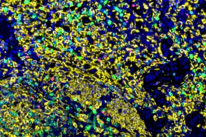
Optimizing IHC Staining through Automation
Roche Diagnostics Ventana Discovery Ultra Autostainer saves time, decreases variation, and increases efficiency for all your immunohistochemistry needs.
See How it Works
Supporting Your Preclinical Research with ISH
With a deep understanding of the complexities of preclinical research and commitment to scientific excellence, we offer multiple services to assist you throughout your preclinical research. Some of our ISH services include:
- Custom probe design services to ensure optimal performance and specificity for your project. Our experts can design and optimize RNAscope probes for a wide range of RNA molecules for any gene and species.
- Assay development to help you enhance your experimental conditions and achieve the best results possible. Our team will work with you to develop customized protocols for their specific research needs.
- Tissue analysis services to help you visualize and analyze gene expression patterns in cells and tissues. Our experts can provide quantitative analysis of gene expression levels and distribution within samples.
- RNAscope technology offers multiplexing capabilities so you can to detect multiple RNA molecules simultaneously within a single sample. This feature enables you to study complex gene expression networks and gain deeper insights into disease mechanisms.
- High sensitivity and specificity through RNAscope probes which have a high binding affinity for target RNA molecules. This allows for highly sensitive and specific detection of gene expression patterns and is useful for detecting low-abundance transcripts and studying gene expression in rare cell populations.
In Situ Hybridization Testing Methods & Capabilities
We offer a wide range of in situ hybridization testing methods to support gene expression analysis in both research and drug discovery settings. Here are some of our key testing methods:
- RNAscope®: A sensitive, specific tissue assay for examining gene expression at the single cell level with spatial morphological context.
- BaseScope™: BaseScope leverages the same platform as RNAscope but expands the detection capabilities, enabling the detection of short RNA target sequences and enabling the differentiation between highly similar sequences.
- And more
In RNA abundance and gene expression studies, no single technique can provide all the answers. When we pair our ISH methods with cell type-specific immunostaining, our team can generate a more complete picture of mRNA and protein expression within specific cell types in tissue sections. These ISH assays can be used to assess tissue responses to various drug treatments for proof-of-principle and/or efficacy and safety evaluations.
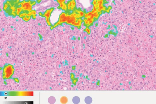
Webinar: Digital Pathology and Image Analysis
Discover the evolution of digital pathology solutions and image analysis, current applications, and future advancements in this expert-led webinar discussion.
Watch Now
Expertise with In Situ Hybridization (ISH) Services
By partnering with us for in situ hybridization services, you will benefit from the expertise, experience, and capabilities of a leading preclinical research organization. With a commitment to customized protocols, multiple detection methods, experienced staff, and comprehensive reporting, we can provide high-quality ISH services to meet your specific needs.
Frequently Asked Questions (FAQs) About In Situ Hybridization Services
-
What is the difference between immunohistochemistry and in situ hybridization?
Immunohistochemistry is the detection of a specific antigen (typically a protein) in thin tissue sections or cells mounted on slides for microscopic evaluation. Immunohistochemistry is the detection of a protein of interest in thin tissue sections or cells mounted on slides for microscopic evaluation. In situ hybridization is also done on thin tissue sections or cells mounted on slides, but it detects a specific sequence or region of DNA or RNA.
-
What makes our ISH services stand out from the competition?
We can work with you closely to develop an immunohistochemistry or immunofluorescence assay specific to your needs. Our scientists have the experience and knowledge needed to understand the nuances of complex projects. Our staining process is automated, resulting in enhanced throughput and consistency.
-
What is a tissue microarray?
A tissue microarray (TMA) consists of multiple small “cores” of tissues on a single microscope slide. Tissue cores are taken from “donor” tissue blocks, which are optimally fixed and preserved tissues taken from healthy animals. TMA blocks can be completely customized with regard to species, tissue types, size, and number of cores.
-
What is tissue microarray technology used for?
Tissue microarray (TMA) slides allow for rapid, high-throughput screening of molecular markers across numerous tissues. It is an efficient, cost-effective way to determine what tissues contain the protein (for IHC) or RNA (for ISH) of interest. TMA slides are also utilized for screening of antibodies during IHC optimization procedures. In this case, tissues are often combined with cell pellets to test specificity and sensitivity of the antibody being tested.