
IHC Services and Techniques
Get your study-specific questions answered with a range of validated molecular pathology procedures, immunohistochemistry techniques, and tailored protocols.
Furthermore, you will have access to tissue microarrays for high-throughput tissue screening of IHC staining. The results can be evaluated by one of our expert pathologists or quantified by automated image analysis.
Advance Your Translational Research with High-Plex Spatial Profiling
This webinar will help you reveal deeper biological insights that accelerate decisions. Discover high‑plex spatial profiling and how it strengthens your translational research.
Watch Now
Common Immunohistochemistry (IHC) Services
- Immune cell profiling/phenotyping and tumor microenvironment characterization
- Track human stem cells xenotransplant into animal models
- Evaluation of reproductive toxicity
- Characterization of brain and nervous tissue cell populations
- Label proliferating cells
- Label apoptotic cells
- Label specific cell types
- Visualizing amyloid plaques in the brain
IHC Testing and Capabilities
- Safety assessments for approximately 70 humanized, chimeric, or murine monoclonal antibodies annually
- Protocol development for over 80 different antibodies in a variety of species
- We have optimized methods for a broad array of markers
- Most of our methods are enhanced for animal tissues, but we are also expanding to include human targets
- All our assays and multiplexes are customizable. If there is a commercially available antibody that has been tested in IHC applications, we can stain for it
- Experience with unconjugated antibodies, antibodies conjugated to a variety of substances, and Fab fragments
- Techniques to reduce or eliminate the binding of secondary reagents to endogenous immunoglobulin, even when the tissue is of the same species as the immunoglobulin
- We use paraffin-embedded and frozen tissue
- Fresh paraffin embedded is most common and is better for multiplexing
- We can also work with frozen and frozen fixed, if your application requires it, but multiplexing options may be limited
- Reproducibility of immunohistochemical techniques in conjunction with quantitative analyses
- QIHC-certified staff using state-of-the-art automated staining instruments
- Roche Ventana Discovery Ultra autostainers
- Hamamatsu s60
- s360 for whole-slide scanning
- Double IHC staining
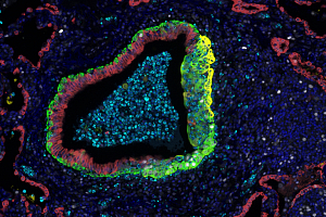
- Single and multiplex chromogenic and immunofluorescence IHC staining with whole-slide imaging for both types of staining
- We can image up to 4 IF channels (typically 3 markers + DAPI nuclear counterstain)

Webinar: How Modern CROs Accelerate Your Drug Development
Are you facing delays, bottlenecks, or rising costs in your drug development process? In this webinar, you will learn how leading CROs overcome these challenges with innovative strategies. Gain insights that can directly impact your timelines, reduce risks, and streamline your path to approval.
Watch the Webinar
Immunohistochemistry vs. In Situ Hybridization
Immunohistochemistry services facilitate the detection of a specific antigen of interest (typically a protein) in thin tissue sections or cells mounted on slides for microscopic evaluation. In situ hybridization is also done on thin tissue sections or cells mounted on slides, but it detects a specific sequence or region of DNA or RNA.
Frequently Asked Questions (FAQs) About Immunohistochemistry (IHC) Services
-
What is immunohistochemistry used for?
IHC services are used when spatial and morphological context of the antigen is desirable. During the preclinical development of novel therapeutics, it is commonly used to assess efficacy or toxicity in vivo. In a clinical setting, it can be used for diagnostic purposes.
-
What makes our IHC services stand out from the competition?
We can work with you closely to develop an immunohistochemistry or immunofluorescence assay specific to your needs. Our scientists have the experience and knowledge needed to understand the nuances of complex projects. Our staining process is automated, resulting in enhanced throughput and consistency.
-
What is a tissue microarray?
A tissue microarray (TMA) consists of multiple small “cores” of tissues on a single microscope slide. Tissue cores are taken from “donor” tissue blocks, which are optimally fixed and preserved tissues taken from healthy animals. TMA blocks can be completely customized with regard to species, tissue types, size, and number of cores.
-
What is tissue microarray technology used for?
Tissue microarray (TMA) slides allow for rapid, high-throughput screening of molecular markers across numerous tissues. It is an efficient, cost-effective way to determine what tissues contain the protein (for IHC) or RNA (for ISH) of interest. TMA slides are also utilized for screening of antibodies during IHC optimization procedures. In this case, tissues are often combined with cell pellets to test specificity and sensitivity of the antibody being tested.