Oncology and Immuno-oncology Assays
Patient-derived xenografts, 3D tumor models, and immune cell co-cultures, these assays replicate the complexity of the tumor microenvironment (TME). This enables accurate assessment of efficacy, immune interactions, and safety, supporting translational success.

AACR Annual Meeting 2026
No matter the circumstances, cancer patients are waiting on new therapies. View our presentations and posters from the AACR Annual Meeting, and browse our resources so you can transform your oncology research with speed and simplicity.
Transform Your Research
Translational PDX Assays
Matched cell line and PDX models enable seamless progression across 2D in vitro, 3D ex vivo, and in vivo assays. With over 1,200 PDX models, 600 commercial tumor cell lines, and a growing organoid bank, screening strategies can be tailored for translational relevance. Proprietary panels include 80 unique cell lines derived from PDX models, characterized for sensitivity to more than 240 reference agents. Assay formats span 3D spheroids, organoids, and 2D immune cell killing platforms, supported by advanced endpoints such as live-cell imaging, cytokine profiling, and genomic analysis.
PDX-Derived Lung Cancer Tumoroids
Tumoroid models provide physiologically relevant 3D systems for evaluating therapeutic potency and immune-mediated cytotoxicity. They replicate tumor complexity and heterogeneity, enabling predictive insights for drug development.
Key Advantages:
- Long-term culture maintains heterogeneous structures for extended studies.
- Biobanking options available (whole tumoroids or suspensions) for future use.
- Size consistency achieved through microwell technologies for reproducibility.
Tumoroid models correlate strongly with in vivo response profiles, making them highly predictive for drug development. They support immuno-oncology studies across hot and cold tumors and allow single-cell characterization using multiplex IHC.
These models deliver quantitative endpoints such as IC50 values for potency comparison and generate sufficient material for OMICS, including genomic and proteomic studies. Their design supports seamless transition from in vitro to in vivo using the same material.
Oncology Preclinical Study Planning Toolkit
Access study pricing, cancer model data, and connect with our oncology experts to plan your oncology efficacy studies with confidence.
Find the Right Model
Dose-Response Profiles of Lung Cancer Tumoroids
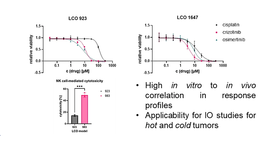
Curves show IC50 values for two tumoroid models (LCO 923, LCO 1647), highlighting predictive correlation between in vitro and in vivo response profiles.
PDX Spheroid Assays
PDX-derived spheroid models replicate cell-cell and cell-matrix interactions, providing a physiologically relevant environment for assessing compound penetration and efficacy. These models provide predictive insights by closely correlating in vitro results with in vivo outcomes.
Key Advantages:
- Mimics physical barriers for realistic drug penetration studies
- Supports potency ranking before costly in vivo studies
- Compatible with molecularly characterized tumor panels
Dose-Response Curves in 3D Spheroid Assay
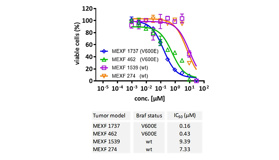
Comparison of sensitivity to Braf inhibitor vemurafenib in melanoma cell lines: V600E mutated vs. wild-type.
PDX spheroid assays provide quantitative endpoints such as IC50 and IC90 values, enabling robust comparison of therapeutic performance. They are ideal for evaluating targeted therapies and immuno-oncology strategies, ensuring translational relevance.
T Cell Killing Assay for Bispecific Antibodies
This assay evaluates the in vitro anti-tumor activity of T cell-engaging antibodies using co-culture systems with human T cells and cancer cell lines. It provides quantitative insights into antibody-mediated cytotoxicity and tumor cell viability.
Key Advantages:
- Works with PDX-derived and publicly available tumor cell lines
- Incorporates LDH release as an additional cytotoxicity indicator
- Enables screening across tumor panels for candidate selection
T Cell-Mediated Cytotoxicity against L363 cells after 24h Co-Culture with Pacanalotamab
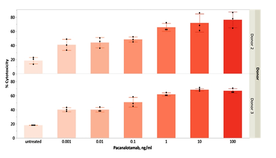
Cytotoxicity (%) measured after 24h co-culture with Pacanalotamab at increasing concentrations (untreated to 100 µg/mL) for two donors (upper bars: Donor 2; lower bars: Donor 3).
Endpoints include flow cytometry for target cell lysis and viability assays for residual tumor survival. These capabilities support comparative evaluation of bispecific antibody performance and streamline candidate preselection for in vivo testing.
Educated CD8+ T Cells
Educated CD8+ T cells are developed to target tumor-specific antigens, enhancing cytotoxic activity against cancer cells. This approach uses peripheral blood mononuclear cells (PBMCs) co-cultured with treated cancer cells and cytokines to prime T cells for tumor-specific killing. Assay read-outs include live cell imaging for PK analysis.
Key Advantages:
- Supports antibody-dependent cell-mediated cytotoxicity (ADCC) studies
- Non-invasive, time-resolved monitoring of immune cell killing
- Compatible with 2D and 3D culture setups
Time-Resolved Killing Activity of Educated CD8+ T Cells in JIMT-1 Model
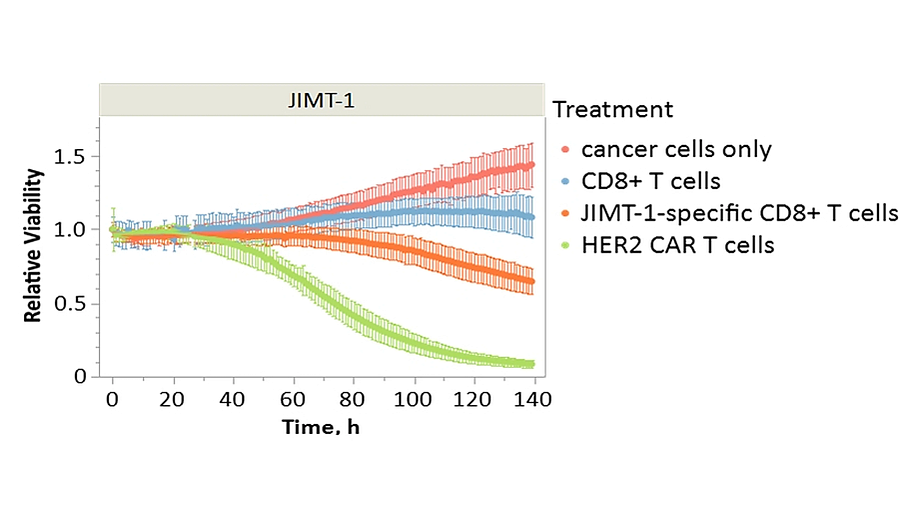
Relative viability of JIMT-1 cancer cells over time when co-cultured with different treatments: cancer cells only, CD8+ T cells, JIMT-1-specific CD8+ T cells, and HER2 CAR T cells.
These assays allow researchers to characterize tumor-specific immune responses, compare the killing efficiency of engineered T cells such as CAR T cells, and generate actionable insights for immuno-oncology development.
Translational Immuno-Oncology Assays
Optimized cancer cell-based assays validate compound efficacy in increasingly complex co-culture assays.
These cancer cell-based assays monitor T cell activation, proliferation, exhaustion, chemotaxis, and cytokine response. as well as interaction with specific cell types such as natural killer cells, macrophages, dendritic cells, neutrophils, and fibroblasts.
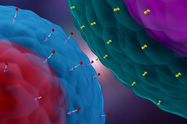
CARrying Your CAR-T to the Clinic
Chimeric Antigen Receptor (CAR)-based therapies are rapidly developing in oncology and other fields. With notable improvements in efficacy, safety, and complexity comes an increased need to streamline the development process.
Watch the Webinar
Have another question or need advice on which model is right for you?
Frequently Asked Questions (FAQs) in Cancer Cell-based Assays
-
What are PDX Assays?
Cells from patient derived xenograft (PDX) are cultured so that oncology therapies can be tested in vitro cancer cell-based assays in material matching the in vivo PDX models, allowing the prediction of efficacy before entering expensive in vivo studies.
-
What is a translational immuno-oncology drug discovery program?
From hit identification to lead validation and in vivo efficacy, a predictive immuno-oncology platform characterizes an immunotherapeutic across the discovery continuum. Using both cancer cell-based assays and in vivo models, the immuno-oncology platform involves integrated approaches toward clinical success of biologics and small-molecule modulators.
-
What are T cell assays?
T cell assays are a type of cancer cell-based assay used to observe the impact of an immunotherapeutic on T cell modulation, such as T cell activation, T cell proliferation, T cell exhaustion, T cell chemotaxis, and cytokine response.
-
Can in vitro immuno-oncology assays help with in vivo model selection?
Yes, in vitro immuno-oncology assays can help with the choice of in vivo model to help mitigate the risk of expensive in vivo pharmacology. For example, cancer cell-based assays, such as PDX-specific tumor killing assays, can be used to select the most relevant PDX in vivo model.
-
How are checkpoint inhibitors used in combination therapies?
Immune checkpoint inhibitors have led to a paradigm shift in the approach to treating a growing list of cancer types. The problem is only about a fifth of patients respond to checkpoint inhibitor monotherapy. Hence, the race is on to boost the response rates by partnering leading checkpoint inhibitors with established therapies (such as chemotherapy) and those which target novel mechanisms. Charles River’s combination screening assays and other cancer cell-based assays can be used to assess your combination therapy strategy.
-
How do you monitor for immunotoxicity?
Immune therapies stimulate the immune system to fight the tumor, however this immune stimulation can have diverse negative immune-mediated side effects. Thus, the increase in immune-oncology drugs and the need to be able to predict and measure potential immunotoxicological consequences is becoming more and more important. Charles River’s cancer cell-based assays and predictive immunotoxicology assays can be used to mitigate this risk.