
EAE Multiple Sclerosis Mouse Models
Experimental autoimmune encephalomyelitis (EAE) multiple sclerosis mouse models are standard models for brain inflammation and demyelination studies. EAE models are induced via injection of myelin oligodendrocyte glycoprotein (MOG) or proteolipid protein (PLP). Disease course and the effect of novel therapeutics are then examined over several weeks. Validated for rats and mice, our EAE models allow you to effectively evaluate novel MS therapies that target the inflammatory response:
- MOG35-55 EAE in C57BL/6 mice
- Spinal cord homogenate (SCH) EAE in rats
- MOG1-125 EAE in Dark Agouti rats
Is the EAE model the best model for multiple sclerosis studies?
Several MS models exist, but by far the best understood and most commonly used is the rodent model of EAE. EAE mouse models are more frequently used because of the inbred genotype of laboratory mice, their rapid breeding capacity, the ease of genetic manipulation and the accessibility of transgenic and knockout mice to facilitate mechanistic studies.
Our EAE Mouse Model Assays
We’ve validated our EAE MS mouse models with positive control compounds, including dexamethasone, glatiramer acetate, and fingolimod. Using a multidisciplinary approach, including translational imaging, behavior and motor function testing, and molecular biomarker analysis, allows the monitoring of both disease progression and therapeutic response in EAE models.
Translational assays that can be used in studies with EAE models include:
- Open field behavior testing for motor function
- Fine motor and gait analysis to examine alterations in parameters like inter-limb coordination, limb movements, and body posture
- MRI with Gadolimium contrast agent to examine and quantify white matter lesions
- SPECT/CT monitoring of inflammation in the brain and spinal cord
- Flow cytometry-based immunophenotyping of peripheral and central nervous system tissues
- Spinal cord and brain pathology monitored via histological techniques and ELISA assays
- Microglial activation assays with EAE model isolated microglia, including phagocytosis and cytokine release assays
- NanoString gene expression profiling on spinal cord or brain tissues, using autoimmune gene profiling panels and pathway analysis to reveal changes in global gene expression
Studies using large animal EAE models include MRI analysis and pathological assessment of lesions.
Imaging in EAE Mouse Model
In vivo imaging in rodent disease models enables the non-invasive investigation of drug efficacy against various pathologies. PET imaging with TSPO ligands, such as 18F-FEPPA, allows for visualization of neuroinflammatory responses, a key pathology and therapeutic target in multiple sclerosis. The data below show the profiling of neuroinflammation by PET imaging in a MOG35-55 EAE mouse model.
MOG35-55 treatment in C57Bl/6 mice induces chronic and progressive disease manifestation from one to two weeks after immunization, with EAE mice showing reduced body weight and higher clinical disease scores compared to controls from 12 days post-immunization. 18F-FEPPA PET imaging reveals increased neuroinflammation across the whole brain, as well as cervical, thoracic and lumbar spinal cord sections 28 days post immunization.
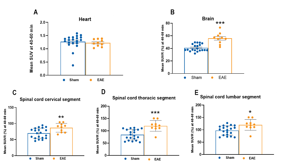
18F-FEPPA activity in the brain (B), spinal cord cervical (C), thoracic (D) and lumbar (E) segments. Heart (A) activity was measured as a control tissue. The data are presented as mean ± SEM of the SUV at 40-60 min post tracer injection (A). For B-E, the data are presented as standardized uptake value ratios (SUVR) between the target region and heart, presented as mean percentages ± SEM. Sham, n=20; EAE, n=10. * p < 0.05, ** p < 0.01, *** p < 0.001 (Welch’s t-test).