
Importance and impact of lysosomal trapping on drug distribution
While a drug’s plasma concentration is the most readily accessible measure of its bioavailability, its disposition – and often its effect – may ultimately depend on free intracellular drug concentrations in the respective tissues. Therefore, understanding the mechanisms that govern cellular unbound drug concentration is essential for reliable in vivo pharmacokinetic and pharmacodynamic predictions based on in vitro data from different systems. In addition to determining unbound drug plasma concentrations and addressing organ-specific uptake rates and partitioning dynamics, intracellular disposition of a compound can also affect its pharmacokinetics. Present in most eucaryotic tissues, lysosomes are not only compartments for cellular deposit, but fundamental elements of physiologic processes such as plasma membrane repair, recycling of cell-surface receptors, reactive oxygen species (ROS) generation, inflammation, cell survival, and cell death1.
Lysosomal trapping or lysosomal sequestration of a compound leads to its accumulation in the lysosomes from the cytoplasm. Such an intracellular distribution limits potential interaction of the compound with a target localized in the cytoplasm or nucleus as well as its availability for metabolic processes, especially in lysosome-rich organs such as the liver or the lungs1. Trapping of a compound can also affect how cellular accumulation data should be interpreted, as in this case the cytoplasmic free fraction of the compound would be much lower than the total intracellular quantities. Lysosomal trapping can therefore have complex implications, including:
- Limited target engagement and efficacy issues2-3, tumor resistance to treatment4
- Effective dose shift and clearance shift2-3
- Drug-drug interactions at the lysosomal level in case of co-administration with other lysosomotropic compounds5
- Adverse effects such as phospholipidosis6 or loss of lysosomal function due to pH shift5
Lysosomal targeting of drugs can also be leveraged as an important strategy either for indications related to lysosomal dysfunction or to leverage lysosome-dependent cell death for cancer therapy7.
Investigation of the lysosomal sequestration of a compound may therefore be relevant in multiple scenarios and phases of drug development. In case lysosomal interactions are either a goal or a considerable risk in the development strategy, early trapping screens can be warranted. As a mechanistic investigation assay, it can also be leveraged to understand potential discrepancies between in vitro and in vivo pharmacokinetic observations. Data on compound unbound fractions in specific cell types and subcellular organs is also often used to improve the predictive performance of in silico PBPK modelling8-11, an approach applied more and more commonly in drug development projects.

How Modern CROs Accelerate Your Drug Development
Are you facing delays, bottlenecks, or rising costs in your drug development process? In this webinar, you will learn how leading CROs overcome these challenges with innovative strategies. Gain insights that can directly impact your timelines, reduce risks, and streamline your path to approval.
Watch the Replay
Sequestration of molecules into the lysosomes (Figure 1A) is driven by the pH difference between the nearly neutral cytosol (pH 7.2) and the acidic lysosomal lumen (pH 4-5), and therefore mostly affects lipophilic basic compounds (~ ClogP>2, pKa 6.5-11). These lipophilic basic compounds can freely diffuse across the phospholipid membrane in their unionized form to enter the cell and the lysosome. In the acidic environment of the lysosome (pH 4–5), the Henderson-Hasselbalch equilibrium shifts toward the compound’s protonated form, which has markedly reduced permeability across lipid bilayers and therefore gets trapped in the lysosome1.
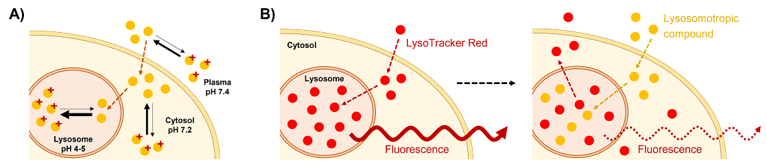
Figure 1. A) Overview of the mechanism of lysosomal trapping. The basic compound (yellow circles) can diffuse from the plasma or medium to the cytosol and then passively enter the lysosome. In this more acidic environment, the equilibrium shifts towards the protonated form of the compound (marked with red +) that has much lower passive permeability and therefore gets trapped in the lysosome. B) Mechanism for detecting lysosomal trapping indirectly based on the extrusion of a fluorescent dye (red circles) from the lysosomal space, leading to a decrease in lysosomal fluorescent signal upon administration of a lysosomotropic compound (yellow circles).
In addition to its impact on overall drug disposition, drugs undergoing lysosomal trapping may also affect sequestration of other lysosomal molecules – endogenous substrates or xenobiotics – and therefore potentially interfere with lysosomal function to some degree. However, there are several other mechanisms that have a broader impact on lysosomes. For example, NH4Cl neutralizes the intraorganellar pH upon its lysosomal uptake, essentially eliminating the pH differences that are a prerequisite for lysosomal trapping of compounds14-16. Such a strong effect on pH is, however, rare among drug candidates. A few other mechanisms may also result in disruption of the lysosomal function, for example interfering with Cyclin G‑associated kinase (GAK) functions affects lysosomal dynamics17, while inhibitors of the vacuolar type
To evaluate lysosomal trapping of drugs, different methods are available. The two most common approaches are:
- A higher-throughput indirect approach with fluorescence-based quantification of changes in lysosomal dye accumulation using high-content imaging (HCI)
- Direct determination of intracellular concentration of a drug via LC-MS/MS in the presence and absence of lysosome inhibitors12
Selection of the lysosomal trapping assay system best suitable for each drug development project depends on multiple factors (see table below). The indirect approach with readouts based on the displacement of a lysosomally accumulated fluorescent dye allows for higher throughput experiments and screening-type approaches thanks to high-content imaging applications. However, while also sensitive, this approach cannot differentiate between dye displacement due to the trapping of another lysosomothropic compound, or other effects on lysosomal function that could reduce dye accumulation. Direct quantification of compound trapping using LC-MS/MS readouts presents a more quantitative approach, allowing direct determination of trapped compound quantities. Other, less widespread methods include application of artificial lysosomal fluid to predict lysosomal trapping of the compounds without cells13.
| Readout type | High-Content Imaging (HCI) | LC-MS/MS |
|---|---|---|
| Principle | Indirect detection of lysosomal sequestration via Lysotracker Red dye extrusion | Direct measurement, % change in accumulation in presence of lysosomal inhibitor |
| Cell types available | MDCKII, Caco-2 or human hepatocytes | Caco-2 or human hepatocytes |
| Positive control |
| |
| Negative control |
| |
| Additional controls |
| |
| Recommended inhibitor | NH4Cl (Ammonium-chloride) | Bafilomycin A1 |
| Recommended application | Earlier stage or preliminary screening - higher throughput, screen-like format available, high sensitivity | Mechanistic investigations - direct measurement of compound lysosomal trapping, lower throughput |
Lysosomal sequestration assessment using high-content imaging (HCI)
For investigating lysosomal trapping of drugs, the most common approach is based on the quantification of changes in lysosomal dye accumulation in the presence and absence of the compound of interest. The LysoTracker Red dye has been specifically developed for visualizing lysosomes as it is strongly lysosomotropic (preferentially accumulates in the lysosomes) and allows for fluorescent detection14. Compounds that are also subject to lysosomal sequestration compete with the dye and their trapping leads to a concentration-dependent decrease of LysoTracker signal in the lysosomes (Figure 1B). Based on this relative signal reduction compared to the dye applied alone, expressed at percent (%) remaining signal, an IC50 value can be calculated to quantify the degree of lysosomal interaction.
Known lysosomotropic compounds, such as chloroquine or propranolol, can be used as positive control for inhibition of lysosomal dye accumulation. It should be noted, however, that other mechanisms exist that interfere with lysosomal function (as described earlier), that may not be differentiated using this indirect approach. Decreased trapping of the LysoTracker dye may also be the result of various potential compound effects on lysosomal function. In most cases, however, dye extrusion due to competitive lysosomal trapping is assumed. If mechanisms other than competitive lysosomal accumulation are suspected, or the degree of sequestration needs to be quantified, direct comparison of test compound accumulation via LC/MS in the presence and absence of lysosomal inhibitors (e.g., Bafilomycin A1) is recommended.
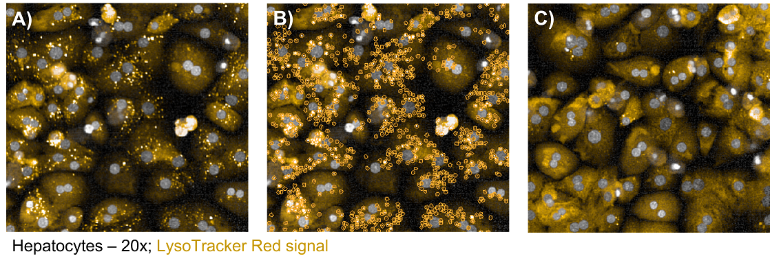
Figure 2. – Cell images of human primary hepatocytes captured by High-Content Imaging (HCI) using 20x magnification air objective. LysoTracker red (LTR) dye (applied at 50 nM) signal is shown in yellow with A) no lysosomal trapping inhibitor added B) lysosomes marked and identified by the HCI image analysis software for fluorescence quantification. C) LTR signal upon treatment with the NH4Cl lysosomal function inhibitor (10 mM).
The fluorescent signal of the LysoTracker dye can be quantified using various detection methods. Leveraging our High-Content Imaging (HCI) platform, considerably higher detection accuracy and sensitivity can be achieved via the traditionally applied microplate reader-based approach. HCI-based cell detection allows for the detection of not only individual cells, but lysosomes as well, so changes in the fluorescent output can be detected precisely at the lysosomal level (Figure 2). As a microplate reader detects signal from a whole well, relatively high background signals are obtained in control conditions where no specific lysosomal trapping occurs. This is, however, almost fully eliminated by applying a HCI approach.
In our assay system, IC50 values of multiple known lysosomotropic drugs, chloroquine, imipramine, and propranolol as inhibitors of the lysosomal trapping of LysoTracker Red were confirmed to be in line with literature findings. Data was found to be highly similar across multiple cell types: MDCKII canine kidney cells, Caco-2 and hepatocytes from both human and rat. Further illustrating the sensitivity of this method, we were also able to determine IC50 values for Verapamil, a compound previously proven to affect lysosomal sequestration of other compounds in the blood-retinal barrier, and Ketoconazole, a drug shown to induce phospholipidosis and predicted to accumulate in lysosomes6, where no IC50 data was previously reported in literature. The improved overall detectability of lysosomal interactions also reduces the chance of generating false negative results.
While inhibition data was similar for the tested cell types, choice of the assay system should be informed by compound characteristics and existing data on its tissue- and sub-cellular distribution. MDCKII, and especially Caco-2 cells, reliably predicted lysosomal trapping and can be used in testing without the additional cost associated with the application of cryopreserved hepatocytes. Cryopreserved hepatocytes, however, express a number of characteristic uptake transporters, including OATP1Bs that are mostly absent in MDCKII, Caco-2, as well as most commercially available immortalized hepatic cell lines, which can contribute to intracellular compound accumulation, allowing a more complex detection of compound handling in case a thorough mechanistic investigation is needed.
Direct lysosomal trapping assay using LC-MS/MS readouts
While for many potential uses, for example to confirm lysosomal targeting where needed or to rule out compound trapping, the identification of lysosomal interaction itself via an indirect approach is sufficient. However, information on the magnitude of lysosomal sequestration may also be necessary. For this, intracellular levels of the test drug itself need to be directly quantified and compared in the presence and absence of a lysosomal function inhibitor such as Bafilomycin A1.
Our assay for detecting lysosomal trapping of compounds directly with LC-MS/MS (or liquid scintillation in case test compounds are [3H] or [14C] labelled), was to set up and optimize in Caco-2 cells and hepatocytes. The reference substrates used for the development of this assay were chloroquine, propranolol, and imipramine. Chloroquine has been retained as the recommended positive control in the assay, as is showed the highest degree of lysosomal accumulation. As a negative control, Rosuvastatin is used in Test compounds, and controls are tested in the presence and absence of a lysosomal function inhibitor.
After incubation with the substrate or test compound with and without inhibitor, the cells are lysed and the effect of the inhibitor on intracellular compound accumulation is quantified. In case a decrease in overall intracellular concentration is observed, the compound is assumed to undergo lysosomal trapping. Data is provided as % compound accumulation in the presence of the inhibitor versus the no-inhibitor control condition which is used for lysosomal trapping classification as “strong”, “weak,” or not trapped:
| Lysosomal trapping | Lysosomal accumulation (%) |
|---|---|
| strong | 50%< |
| weak | 20%-50% |
| none | <20% |
Both NH4Cl and Bafilomycin A1 were considered as potential reference inhibitors in this assay system. Historically, NH4CI has been more extensively used in lysosomal trapping experiments, therefore more literature data is available with this compound. However, upon completion of comparative tests using all three above-listed probe substrates, we determined that in our setup and all tested cell types, the use of Bafilomycin A1 leads to improved fold-change detection of trapped compounds over control conditions where no inhibitor is applied. Therefore, we apply Bafilomycin A1 as reference inhibitor to improve sensitivity of our lysosomal trapping assays with LC-MS/MS readout (for HCI-based assays, NH4Cl will remain the recommended inhibitor, as it does not require preincubation with the cells and is thus more compatible with this quick and sensitive screen-type assay).
-
References
1MacIntyre, A. C., & Cutler, D. J. (1988). The potential role of lysosomes in tissue distribution of weak bases. Biopharmaceutics & drug disposition, 9(6), 513–526. https://doi.org/10.1002/bod.2510090602
2Daniel, W. A., & Wójcikowski, J. (1999). Lysosomal trapping as an important mechanism involved in the cellular distribution of perazine and in pharmacokinetic interaction with antidepressants. European neuropsychopharmacology: the journal of the European College of Neuropsychopharmacology, 9(6), 483–491. https://doi.org/10.1016/s0924-977x(99)00034-6
3Llanos, S., Megias, D., Blanco-Aparicio, C., Hernández-Encinas, E., Rovira, M., Pietrocola, F., & Serrano, M. (2019). Lysosomal trapping of palbociclib and its functional implications. Oncogene, 38(20), 3886–3902. https://doi.org/10.1038/s41388-019-0695-8
4Hraběta, J., Belhajová, M., Šubrtová, H., Merlos Rodrigo, M. A., Heger, Z., & Eckschlager, T. (2020). Drug Sequestration in Lysosomes as One of the Mechanisms of Chemoresistance of Cancer Cells and the Possibilities of Its Inhibition. International journal of molecular sciences, 21(12), 4392. https://doi.org/10.3390/ijms21124392
5Logan, R., Funk, R. S., Axcell, E., & Krise, J. P. (2012). Drug-drug interactions involving lysosomes: mechanisms and potential clinical implications. Expert opinion on drug metabolism & toxicology, 8(8), 943–958. https://doi.org/10.1517/17425255.2012.691165
6Hanumegowda, U. M., Wenke, G., Regueiro-Ren, A., Yordanova, R., Corradi, J. P., & Adams, S. P. (2010). Phospholipidosis as a function of basicity, lipophilicity, and volume of distribution of compounds. Chemical research in toxicology, 23(4), 749–755. https://doi.org/10.1021/tx9003825
7Groth-Pedersen, L., Aits, S., Corcelle-Termeau, E., Petersen, N. H., Nylandsted, J., & Jäättelä, M. (2012). Identification of cytoskeleton-associated proteins essential for lysosomal stability and survival of human cancer cells. PloS one, 7(10), e45381. https://doi.org/10.1371/journal.pone.0045381
8Bolger, M. B., Macwan, J. S., Sarfraz, M., Almukainzi, M., & Löbenberg, R. (2019). The Irrelevance of In Vitro Dissolution in Setting Product Specifications for Drugs Like Dextromethorphan That are Subject to Lysosomal Trapping. Journal of pharmaceutical sciences, 108(1), 268–278. https://doi.org/10.1016/j.xphs.2018.09.036
9Wu, D. N., Yu, G., & Li, G. F. (2022). Reproducing prolonged time to peak bisoprolol concentration by PBPK modeling with lysosomal trapping in enterocytes. CPT: pharmacometrics & systems pharmacology, 11(1), 5–6. https://doi.org/10.1002/psp4.12756
10Liu, X., & Jusko, W. J. (2021). Physiologically Based Pharmacokinetics of Lysosomotropic Chloroquine in Rat and Human. The Journal of pharmacology and experimental therapeutics, 376(2), 261–272. https://doi.org/10.1124/jpet.120.000385
11Bauch, C., Bevan, S., Woodhouse, H., Dilworth, C., & Walker, P. (2015). Predicting in vivo phospholipidosis-inducing potential of drugs by a combined high content screening and in silico modelling approach. Toxicology in vitro : an international journal published in association with BIBRA, 29(3), 621–630. https://doi.org/10.1016/j.tiv.2015.01.014
12Schmitt, M. V., Lienau, P., Fricker, G., & Reichel, A. (2019). Quantitation of Lysosomal Trapping of Basic Lipophilic Compounds Using In Vitro Assays and In Silico Predictions Based on the Determination of the Full pH Profile of the Endo-/Lysosomal System in Rat Hepatocytes. Drug metabolism and disposition: the biological fate of chemicals, 47(1), 49–57. https://doi.org/10.1124/dmd.118.084541
13Yousef, Malaz, et al. "Sub-cellular sequestration of alkaline drugs in lysosomes: new insights for pharmaceutical development of lysosomal fluid." Research in Pharmaceutical Sciences 18.1 (2023): 1. https://doi.org/10.4103/1735-5362.363591
14Chazotte B. (2011). Labeling lysosomes in live cells with LysoTracker. Cold Spring Harbor protocols, 2011(2), pdb.prot5571. https://doi.org/10.1101/pdb.prot5571
15Hart, P. D., & Young, M. R. (1991). Ammonium chloride, an inhibitor of phagosome-lysosome fusion in macrophages, concurrently induces phagosome-endosome fusion, and opens a novel pathway: studies of a pathogenic mycobacterium and a nonpathogenic yeast. The Journal of experimental medicine, 174(4), 881–889. https://doi.org/10.1084/jem.174.4.881
16Ling, H., Ardjomand, P., Samvakas, S., Simm, A., Busch, G. L., Lang, F., Sebekova, K., & Heidland, A. (1998). Mesangial cell hypertrophy induced by NH4Cl: role of depressed activities of cathepsins due to elevated lysosomal pH. Kidney international, 53(6), 1706–1712. https://doi.org/10.1046/j.1523-1755.1998.00952.x
17Miyazaki, M., Hiramoto, M., Takano, N., Kokuba, H., Takemura, J., Tokuhisa, M., Hino, H., Kazama, H., & Miyazawa, K. (2021). Targeted disruption of GAK stagnates autophagic flux by disturbing lysosomal dynamics. International journal of molecular medicine, 48(4), 195. https://doi.org/10.3892/ijmm.2021.5028
18Fedele, A. O., & Proud, C. G. (2020). Chloroquine and bafilomycin A mimic lysosomal storage disorders and impair mTORC1 signalling. Bioscience reports, 40(4), BSR20200905. https://doi.org/10.1042/BSR20200905