
Mast Cells in Allergies and Other Inflammatory Conditions
Mast cells (MCs) are granulated, long-living innate cells found in peripheral tissues such as skin, airways, and gastrointestinal tract. They are the first responders to allergens and parasitic infections. Classically, mast cells are activated by the binding of the FɛRI (the high affinity surface receptor for IgE) to an antigen-bound IgE-sensitized cell. A second, non-classical mechanism is driven by the activation of surface molecules such as C5aR or MRGPRX2, which contribute to multiple inflammatory conditions, including neurogenic inflammation. Upon activation, mast cells release a wide range of inflammatory and vasoactive mediators such as histamine, proteases (e.g., tryptase, chymase), leukotrienes (e.g., LTC4) prostaglandins (e.g., PDG2) and lysosomal enzymes (e.g., β-hexosaminidase) as well as they upregulate CD63, a key activation biomarker. This rapid process, called degranulation, leads to vascular changes, inflammation, and to the recruitment of other immune cells, which contribute to the development of itching, coughing, and sneezing and in some cases to severe life-threatening conditions.
In vitro MCs assays are a valuable tool for the profiling of therapeutics, that inhibit or stabilize or modulate MCs activation for the treatment of MC-mediated diseases.
Human MCs are differentiated from human CD34+ cells cultured for ~ 6-8 weeks in the presence of hIL-3, hIL-6 and hSCFC (Stem cell Factor/cKit ligand). Differentiated MCs are characterized by the cell surface expression of c-Kit/CD117, a transmembrane tyrosine kinase which is a key regulator for MCs proliferation and survival and can be triggered with either IgE/anti-IgE-antibody or IgE-independent triggers (e.g., substance P or C5a) for functional assays.
Immunology Brochure
Browse our immunology brochure to learn more about our wide range of capabilities from target discovery to early clinical. Our facilities in North America and Europe can support your drug discovery journey through every step of the workflow.
Download now
Key Assays to Target Mast Cell Function
Charles River has developed in vitro MCs differentiation and activation assays for the screening of therapeutics targeting MCs function. Below are listed the main assays that Charles River can perform with MCs:
- MC Differentiation: Human CD34+ progenitors isolated from healthy donors are cultured for ~ 6-8 weeks in the presence of cytokines and growth factors that promote mast cell differentiation and expansion. This process generates a high number of MCs for therapeutic candidate screening, which would not be sufficient when using tissue-resident MCs.
- MC Immunophenotyping: Upon differentiation, MCs are characterized by flow cytometry to assess the expression of MC lineage biomarkers (e.g., c-kit/CD117 , FcεRI), IgE-independent biomarkers (e.g., MRGPRX2, C5aR) as well as activation biomarkers (e.g., CD63) by flow cytometry.
- MC Activation and Degranulation: Differentiated MCs are activated in vitro with soluble triggers (e.g., IgE/anti-IgE-antibody, Substance P, C5a) to induce MC degranulation which releases soluble mediators (e.g., Tryptase, β-hexosaminidase) and upregulates surface biomarkers (e.g., CD63). Plate readers-based kits are used for the detection of soluble mediators, while flow cytometry is used to assess surface biomarker expression. Drug candidates are added to assess their capability to modulate MCs function.
Mast cells are effector cells playing a key role at the edge between the environment and the host. In vitro mast cell assays are valuable tools to evaluate all stages of mast cell-associated diseases. Charles River provides in vitro mast cell assays for determining MoA or efficacy of mast cell-targeting therapeutics.
Representative Data
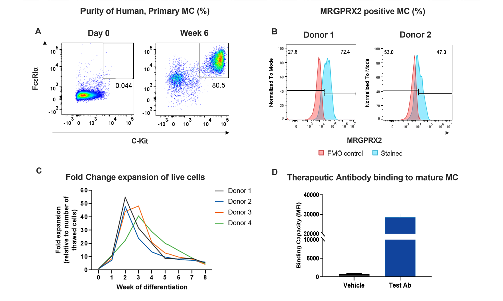
Figure 1. Human Primary MCs differentiation and immunophenotyping. MCs are differentiated from CD34+ cells (Hematopoietic Stem cells) for 6-8 weeks in the presence of cytokines/growth factors. A. Surface expression of MC biomarkers (FcεRIα and c-Kit) after differentiation by flow cytometry. B. MRGPRX2 expression in two human MC donors (FcεRIα+ c-Kit+). C. Fold change expansion of MCs during the in vitro differentiation from CD34+ cells from 4 donors. D. Binding of a therapeutic antibody to mature MCs.
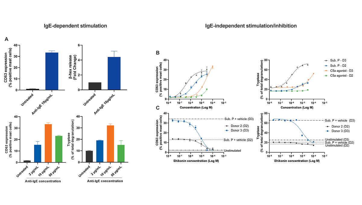
Figure 2. IgE-dependent and IgE-independent MC activation and therapeutic screen. Differentiated human MCs from 2 donors were sensitized with IgE and then treated with anti-IgE to induce degranulation through IgE cross-linking. A. CD63 expression by flow cytometry as well as β-hexosaminidase (β-hex, upper right panel) or tryptase release were measured as readouts of MC activation and degranulation upon different concentration of anti-IgE (lower panel). B. IgE-independent MC activation of 2 donors measured by CD63 expression and tryptase release upon stimulation with substance P or C5a agonist. C. Shikonin-mediated inhibition (IgE-independent activation) of MC activation and degranulation measured by CD63 expression and tryptase release upon substance P stimulation on 2 donors.
In addition to in vitro mast cell assays, Charles River also provides in vivo mast cell models, such as Asthma models.
Frequently Asked Questions (FAQs) About Mast Cell Assays
-
What is the role of Mast Cells (MCs)?
Their primary function is to defend our body mucosae and peripheral tissues from parasitic infections and allergens. However, their rapid activation in the tissues can lead to severe allergies, anaphylaxis and autoimmune diseases resulting detrimental triggers of chronic inflammation. MCs stabilizing drugs or inhibitors are commonly used as treatment.
-
How do pathogens activate Mast cells (MCs)?
The most classical mechanism for MCs activation occurs when the high-affinity (IgE) receptor FcεRI expressed on MCs surface binds an antigen-bound IgE. This results in the FcεRI clustering, which in turn induces downstream signaling and consequential release of inflammatory mediators. Parasitic infections from helminths, nematodes, and protozoa are the most frequent triggers of MCs activation, alongside food or skin allergens.
-
What is Mast cells (MCs) lifespan?
MCs are long-lived cells which can survive up to 12 weeks in the skin. They can perform multiple rounds of degranulation and accumulate in organs based on the disease type.
-
How similar or different are Mast cells (MCs) to Basophils?
Both MCs and basophils are crucial players in allergies and inflammatory conditions, and they are a unique source of histamine for our body. Both cell types rapidly degranulate upon high-affinity IgE receptor cross-linking. They differ for tissue distribution (with MCs being tissue-resident cells and basophils being circulating cells) and for their ability to release inflammatory mediators. Histamine and lipid mediators are mediators of both MCs and basophils, while tryptase and other proteases are more specific for MCs. Additionally, they vary in the development process, life-span and pharmacological heterogeneity, which are all relevant factors in the drug development process.
-
Can we use already differentiated Mast cells (MCs) by isolating them directly from the skin?
Despite the substantial improvement in laboratory techniques, purification of human tissue-resident MCs still represents a big challenge and is not commonly used, due to the low cell yield and the impossibility of obtaining large pieces of biopsies. Thus, a differentiation from a cell precursor is the most largely used method to evaluate MCs responses.
-
Do Mast cells (MCs) have other functions besides their role in the host defense and allergies?
Many studies demonstrated that MCs also participate in wound healing and bone remodeling, can help degrading extracellular matrixes and modifying lipoproteins during fibrotic processes.