
What Are Neutrophils?
Neutrophils are potent effector cells of the innate immune system that participate in a wide array of immunological and inflammatory responses. They differentiate and mature in the bone marrow from a granulo-monocytic progenitor (GMP) in response to granulocyte colony-stimulating factor (G-CSF), a critical mediator of their maturation and release into the blood stream.
Neutrophils are often the first line of defense against microorganisms and protect the body by rapidly releasing antimicrobial molecules such as reactive oxygen species (ROS), myeloperoxidases (MPOs), and other hydrolytic enzymes. Their rapid recruitment to the site of inflammation is mediated by chemoattractants detected via their expression of chemokine receptors, formyl-peptide receptor 1, and leukotriene receptors. To eliminate pathogens, neutrophils can also form a web-like complex of chromatin and granule proteins, called neutrophil extracellular traps (NETs). These lead to a unique form of programmed cell death called ‘NETosis,’ which traps pathogens, preventing their spread.
Neutrophils in Diseases and Drug Discovery
Increasing evidence supports neutrophil function in the pathogenesis of a wide range of pathologies such as infections, pulmonary diseases, autoimmune and inflammatory diseases, and cancer. Nevertheless, neutrophil involvement in disease seems to depend on several factors, including aging, disease stage, and microbiome composition. Hence, therapeutic approaches targeting neutrophil development, maturation, function, and migration during different pathological settings are of high interest in the fields of immunity, inflammation, and cancer.
Charles River offers multiple in vitro neutrophil assays to assess the effects of your therapeutics on neutrophil function and accelerate the development of your clinical candidate.
How can we support your program?
Key Neutrophil Assays to Target Neutrophil Function
Charles River has developed in vitro neutrophil assays in both human and rodent species. Neutrophils are isolated from blood samples or bone marrow (rodent) and analyzed by flow cytometry to assess the expression of lineage surface biomarkers. Cells are then immediately used for in vitro functional studies. Shown below is a list of the assays we can perform with neutrophils:
Isolation and Phenotyping
- Neutrophil isolation: Neutrophils can be isolated from buffy coats of healthy donors via Ficoll separation and dextran-based sedimentation, or via the use of commercially available granulocyte-enriching density gradients.
- Neutrophil phenotyping: Isolated neutrophils are analysed by flow cytometry for the expression of neutrophil lineage biomarkers (e.g., CD11b, CD66b, and CD15).
Functional assays
- Neutrophil oxidative burst assay (ROS): Isolated neutrophils are seeded and activated with stimuli (e.g., E. coli, fMLP, PMA) in the presence of a drug candidate. Intracellular ROS production by neutrophils is detected using fluorogenic dye (dihydrorhodamine — DHR 123), which is oxidized in the presence of reactive oxygen species. Myeloperoxidase is measured in the supernatant using an ELISA kit.
- Phagocytosis assay: Isolated neutrophils are incubated with E. coli-FITC for 30 minutes and fluorescence intensity is measured by flow cytometry or high-content analysis (HCA) to assess phagocytic activity.
- CD62L shedding: The amount of CD62L (L-selectin) shed from the surface of neutrophils can be used to measure neutrophil activation or ‘priming.’ Isolated neutrophils are stimulated with fMLP or LPS in the presence of a drug candidate. PMA can be used as positive control. Levels of surface CD62L are then determined by flow cytometry.
- Chemotaxis assay: Isolated neutrophils are seeded in the upper chamber of a 96-well 5 Boyden chamber (5 μm pore-size) in serum-free medium. The chemoattractant of interest (e.g., IL-8) and drug candidate are added to the lower chamber. After 1 hour, neutrophil migration into the lower chamber is quantified by total cellular ATP levels via a luminescent-based method (CellTiter-Glo®).
- NETosis assay: Isolated human neutrophils are seeded and stimulated with either PMA or BSA-Immune Complexes (IC) in a 96-well plate format. Drug candidates and NETosis inhibitor reference compounds (e.g., R406 — Syk-inhibitor Fostamatinib; DPI — ROS-inhibitor) are then added for 3 hours. NETosis is measured by high-content analysis using NUCLEAR ID red and SYTOX Green staining to measure in real-time the loss of cell membrane integrity via a progressive increase in fluorescence intensity combined with NETs spreading beyond cell borders.
We offer multiple in vitro neutrophil assays to assess the effects of your therapeutics on neutrophil function or migration that will accelerate the development of your clinical candidate.
Representative Data
Neutrophils Immunophenotyping and Activation (Oxidative burst)
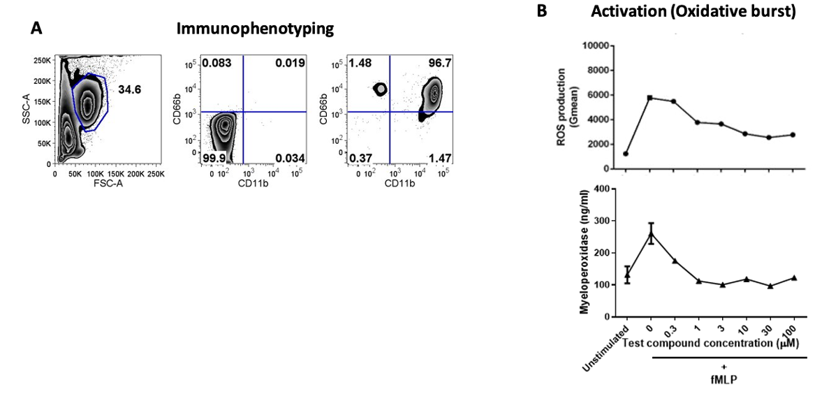
Figure 1. A. Human neutrophils isolated from healthy blood donors were immunophenotyped for lineage surface markers (CD66b, CD11b) by flow cytometry. B. Isolated neutrophils are stimulated with fMLP (N-Formylmethionyl-leucyl-phenylalanine) and in the presence of a test compound at 6-point CRC. B. Compound inhibitory effects are assessed by measuring ROS (Reactive oxygen species) and Myeloperoxidase by commercial kits.
Chemotaxis Assay
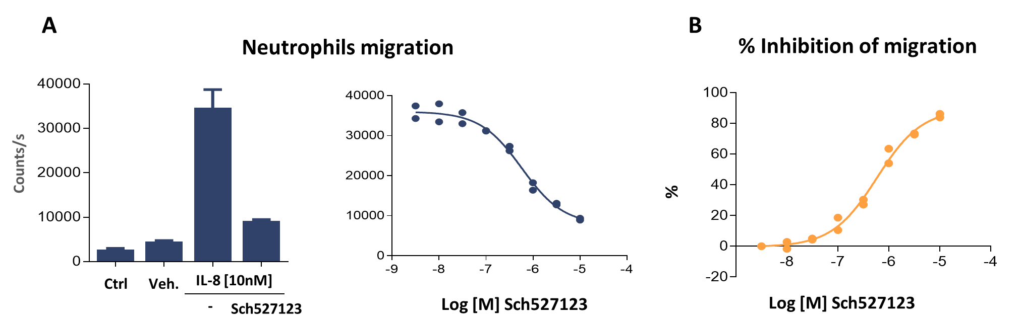
Figure 2. A. Human neutrophils isolated from healthy blood donors were seeded in the upper chamber of a 96-well Boyden chamber that contain 5.0 μm pore-size polyester membrane in serum-free medium, while chemoattractant (IL-8 10nM) and/or compounds are added to the lower chamber. After one hour, neutrophil migration through the pores into the lower chamber is detected by measuring ATP levels via a luminescence-based method (CellTiter-Glo®). B. Data shows that IL-8-mediated migration of human neutrophils is strongly inhibited by treatment with a CXCR1/2 antagonist (Sch527123). Ctrl = Controls; Veh. = Vehicle
NETosis Assay
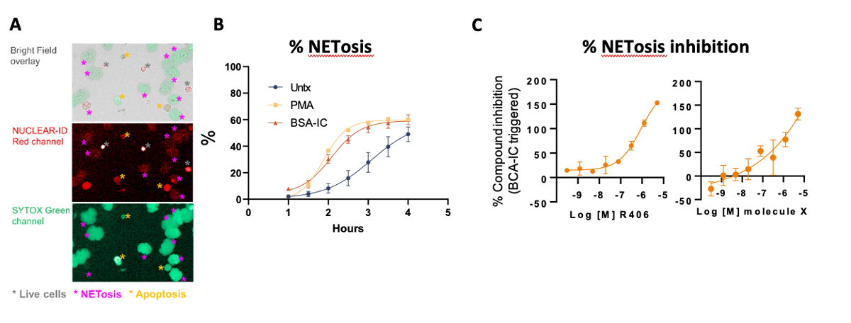
Figure 3. A Representative high-content images of neutrophil nuclear and cytoplasmic changes during different types of cell death (20x magnification). Top, bright field images overlay with NUC with NUCLEAR-ID Red and SYTOX Green channels. Middle, NUCLEAR-ID Red channel. Bottom, SYTOX Green channel. Grey asterisks indicate live cells, magenta asterisks indicate NETotic neutrophils, and yellow asterisks indicate apoptotic neutrophils. B. Percentage of neutrophils undergoing NETosis induced by various stimuli over time. Neutrophils were either left unstimulated or triggered with PMA or BSA-IC. Images were acquired 1 hour post stimulation and then at 30 min intervals. Untx, untreated; IC, Immune complex. C. Representative data of dose-dependent NETosis modulator screening. Both reference compound R406 (left) and testing molecule X (right) showed a dose-dependent inhibition on BSA-IC-induced NETosis. Data are presented as mean ± SD.
Development of In Vitro Assays for the Assessment of Potential Drugs Regulating the Migration of Blood-derived Human Monocytes or Neutrophils
Understanding the switches that regulate migration of innate cells in pathological settings can be a fundamental approach in the fields of inflammation, autoimmune disease, and cancer research.
Download The Poster
Frequently Asked Questions (FAQs) About Neutrophil Assays
-
What is a neutrophil’s lifespan?
Neutrophils have a very short lifespan in the bloodstream. In humans, their circulating half-life is less than 8 hours, while ex vivo their half-life is usually less than 24 hours.
-
How do neutrophils become activated?
Neutrophils are exquisitely sensitive to any experimental manipulation and are hard-wired to respond to many microbial and inflammatory stimuli. Once the body is infected with a pathogen, neutrophils quickly accumulate at the site of inflammation or infection, where they exert their effector functions. This activation leads to the eradication of the invading pathogen via phagocytosis, generation of ROS and granule-derived myeloperoxidase (MPO), hydrolytic enzymes (e.g., elastase, matrix metalloproteinases), and other antimicrobial proteins/peptides (e.g., defensins).
-
How can we target or manipulate neutrophil function in drug discovery?
Several therapeutic approaches that enhance neutrophil activation or production, or promote their inhibition and apoptosis, or even depletion, are currently under investigation for indications such as asthma, rheumatoid arthritis, metastatic melanoma, and type 2 diabetes. These approaches mainly focus on the use of small molecules, biologics, or neutrophil-derived molecules such as granule proteins.