How Can Dendritic Cell Function Be Modified to Help Treat Autoimmune and Inflammatory Diseases?
When exposed to inflammation or pathogen-associated molecular patterns (PAMPs), dendritic cells (DC) are activated and migrate towards lymphoid organs through which naïve T cells circulate. Activated (mature) dendritic cell express high levels of costimulatory molecules and release cytokines which co-ordinate with antigen presentation via MHC to activate CD4 and CD8 T cells, initiating or reactivating an existing immune response.
These pathways play a critical role in protective immunity to pathogens or tumors yet can be dysfunctional in autoimmune disease. As such, there is considerable scope to target various aspects of dendritic cell function therapeutically.
Key Assays to Target Dendritic Cell Function
Charles River has developed a range of in vitro dendritic cell assays which enable characterization of dendritic cell phenotype and function to evaluate the effect of therapeutics in a variety of disease settings:
- MLR assays: co-culturing dendritic cells with allogeneic T cells allows assessment of the effect of a therapy on dendritic cell phenotype and function. This is assessed using flow cytometric analysis, proliferation of T cells, and production of cytokines as readouts.
- By feeding dendritic cell with a tumor-associated antigen or an autoantigen associated with autoimmune responses, we have also developed sophisticated systems to screen for both antigen and therapeutic specificity, and can evaluate the ability of dendritic cell to drive either CD4 or CD8 T cell responses.
- By transducing primary dendritic cell with a bespoke lentivirus containing the protein or antigen relevant to the disease or autoimmune condition, antigen-specific T cells can be expanded even when endogenous expression levels are low or absent in dendritic cell.
- Tolerogenic dendritic cells are important in driving regulatory T cell (Treg) development and differentiate into this phenotype in the presence of high levels of IL-10 or certain bacterial components. Our models can drive the differentiation of this dendritic cell subset in vitro and the ability of a therapeutic to drive a similar polarization can be assessed; readouts include the ability of these tolerogenic cells to alter CD4 T cell polarisation. This readout is specifically targeted for evaluating therapeutics for autoimmune conditions, such as colitis, Crohn’s disease, or rheumatoid arthritis.
Working With Dendritic Cell Assays In Vitro
Dendritic cells comprise three main subsets, conventional (cDC); types 1 and 2, and plasmacytoid (pDC). These cells are identified by their characteristic surface markers and can be isolated in small numbers from the blood to test the effect of therapeutics on d differentiation and function (Figure 1).
Alternatively, when larger numbers of dendritic cell are required, these can be differentiated from monocytes. Immature monocyte-derived dendritic cell (moDC) can be matured by exposing them to a variety of stimuli which in turn dictate the phenotype of the matured cell (Figure 2). This assay can be used to screen infection targeted therapeutics for their ability to promote immature moDC activation, whereas therapeutics that suppress this response in the presence of activating stimuli may be autoimmune targeted.
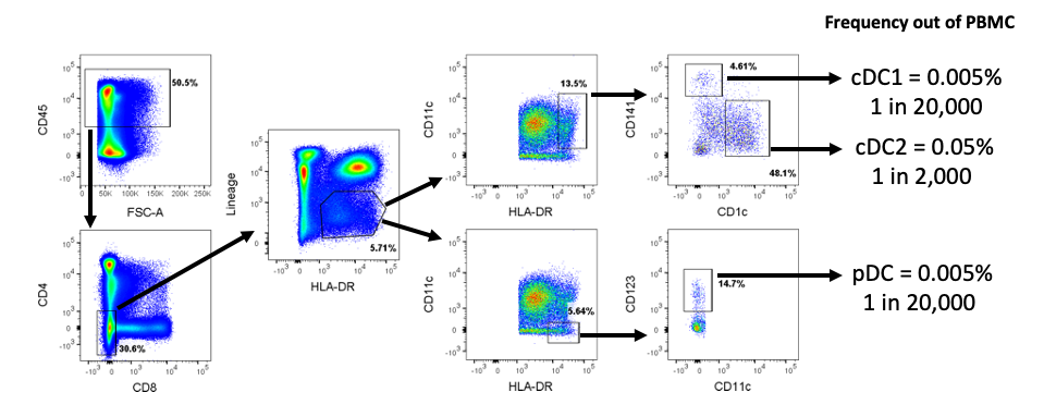
Figure 1: Assessment of dendritic cell subset frequency in peripheral blood mononuclear cells (PBMC) by flow cytometry. A representative gating strategy is shown for the identification of dendritic cell subsets from healthy human PBMC. From single viable cells, gates for CD45+, CD4-, CD8-, Lineage-, and HLA-DR+ were used to identify dendritic cell. Within the dendritic cell population, cDC1 were identified as CD11c+ CD141+ CD1c-, and cDC2 were identified as CD11c+ CD141- CD1c+. pDC were identified as CD11c- HLA-DR+CD123+. The frequency of each subset within total PBMC is shown on the right-hand side of the figure.
Dendritic Cell Assays: Phenotyping
-
Flow cytometry analysis for dendritic cell assays
The ability of therapeutics to alter the activation profile of moDC can be evaluated using flow cytometry.
The addition of LPS to moDC results in their maturation, as evidenced by upregulation of both MHC Class I and II molecules and costimulatory or activation markers such as CD86, CD83, CD80, and CD40 (Figure 2). These phenotypic changes are known to enhance the ability of moDC to stimulate a T cell response and therefore can be used as a surrogate for predicting dendritic cell function.
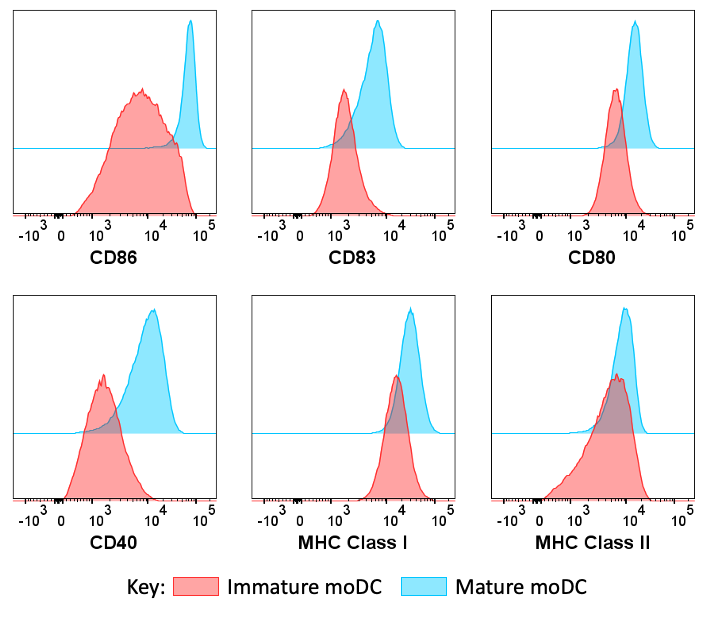
Figure 2: Ddendritic cell assay: phenotypic profiles of LPS-matured and immature moDC by flow cytometry. Histograms show the raw flow cytometry data for surface marker expression from 6 day differentiated moDC, cultured overnight as immature dendritic cell (red) or matured with LPS + IFNγ (blue).
Therapeutics that induce a similar change in the dendritic cell maturation profile are likely to show efficacy in an infection setting while those that block the LPS-induced changes are likely to be suitable for autoimmune disease intervention.
-
Confocal imaging analysis for dendritic cell assays
Confocal imaging or HCS can provide both a greater understanding of mechanism of action and evaluate the sub-cellular localization of targets of interest.
For the example data shown in Figure 3, moDC have been co-cultured with fluorescently labelled IgG as a surrogate for immune complex uptake. The moDC endocytose the IgG which then partially co-localizes with LAMP1, a lysosomal marker, suggesting that the IgG has entered the lysosomal degradation pathway. This uptake mechanism could therefore drive responses to self or pathogenic antigens depending on the disease context.
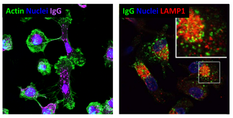
Figure 3: Uptake of immunoglobulin by moDC and sub-cellular localization. Images taken of moDC exposed to fluorescently labelled IgG. On the left-hand side the cells were stained with Actin (green), IgG (purple), and nuclear stain (blue). On the right-hand side, the cells were stained with IgG (green), LAMP1 (red), and nuclear stain (blue). These images show that IgG is internalized by moDC and subsequently partially co-localizes with lysosomes (LAMP1+).
Functional Dendritic Cell Assays
There are several ways to probe the functionality of moDC using in vitro assays.
-
Dendritric Cell maturation cytokine profiles
LPS treatment of moDC induces both inflammatory cytokines such as IL-12, IL-6, and TNFα, and the anti-inflammatory cytokine, IL-10 (Figure 4). The absolute quantities of the cytokines can be used as a functional readout as they will influence the nature of the T cell response.
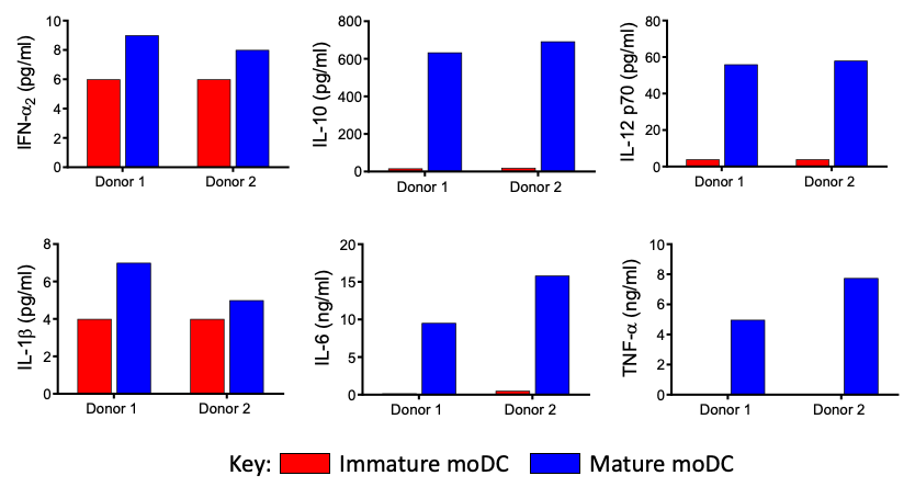
Figure 4: Cytokine production by moDC. moDC were cultured overnight with (blue bars) and without (red bars) LPS. The production of cytokines (IFNα, IL-1β, IL-10, IL-6, IL-12p70, TNFα) were then quantified from supernatants by Luminex.
-
Functional Dendritic Cell and T cell co-cultures
T cell activation can be used as a readout for modulation of dendritic cell antigen presenting function.
MLR assays using allogenic CD4 T cells co-cultured with therapeutically targeted dendritic cell provide a straightforward screening assay to assess dendritic cell antigen presenting function. Readouts of dendritic cell function include T cell proliferation and cytokine production as well as phenotyping of the dendritic cell and T cells by flow cytometry.
The use of mature moDC enhances the ability of CD4 T cells to proliferate and is accompanied by upregulation of MHC Class II and ICAM on the dendritic cells (Figure 5A). In addition, inflammatory cytokines including TNFα, IL-6, and IL-12 go from undetectable levels to a robust response upon maturation of dendritic cell (Figure 5B).
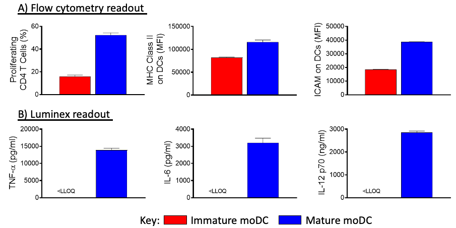
Figure 5: DC and CD4 T cell MLR comparing immature and mature dendritic cell function. The moDC were cultured with and without dendritic cell maturation conditions (LPS + IFNγ). Allogenic CD4 T cells were added for several days before measuring cytokines by Luminex and CD4 proliferation by flow cytometry.
This offers a platform for testing therapeutics that will modify the interaction between the dendritic cell and T cells to either increase, decrease, or repolarize the T cell response.
-
Tolerogenic dendritic cells
Agents that induce a tolerogenic dendritic cell profile can be assessed as demonstrated below
Levels of IL-10 in the culture supernatant were increased in cultures where the therapeutically modulated dendritic cell were present, which could be desirable for the treatment of autoimmune disease.
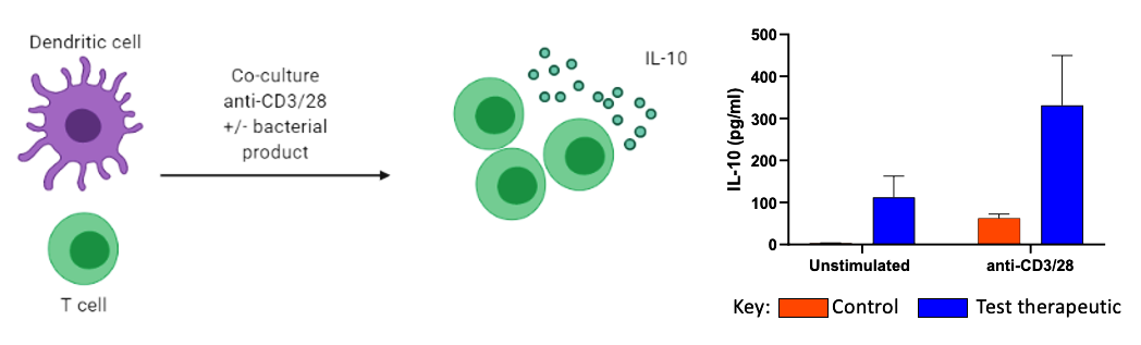
Figure 6: Tolerogenic dendritic cell generation and induction of IL-10-secreting T cells. Monocytes were differentiated into moDC in the presence or absence of a bacterial product. Following this the different moDC populations were co-cultured with autologous naïve CD4+ T cells in the presence of anti-CD3/28 antibodies. Levels of IL-10 secretion in co-cultures were determined by Luminex.
Levels of IL-10 can then be quantified in the culture supernatant, and were increased in cultures where the therapeutically modulated dendritic cell were present, suggesting the treatment could have efficacy in autoimmune disease.
Specialist Dendritic Cell Assays
-
Genetic manipulation of primary moDC
Using nucleofection with a plasmid or mRNA, lentiviral transduction, or siRNA transfection, genetic manipulation can be used to overexpress antigens or to target specific genes associated with dendritic cell function.
For instance, introducing an antigen associated with autoimmune responses that is not typically expressed by dendritic cell allows endogenous processing and presentation to T cells to test modulation of immunogenicity in a range of sophisticated assays.
Genetically modified dendritic cell assay can be combined with any other assay platform to offer a bespoke system, targeting a gene of interest and/or therapeutic.
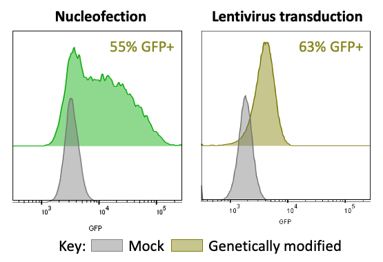
Figure 7: Dendritic cell assay: genetic modification of DC. moDC were subjected to nucleofection with GFP mRNA or lentiviral transduction with a GFP vector. The efficiency of transfection/transduction was determined two days later by flow cytometry compared to mock transduced cells.
-
Human dendritic cell activation of autologous antigen specific T cells
Naïve CD8 T cells can be co-cultured with matured autologous moDC alongside an antigen of interest. This antigen could be a known target, such as a tumor-associated antigen (TAA), an autoimmune antigen, or even a novel target to test its immunogenicity.
The example data here shows a TAA inducing the strong expansion of antigen specific T cells following a two-week co-culture with moDC (Figure 8).
This platform can therefore be utilized to test the efficacy of a compound in the context of rare IO or auto-antigen specific T cell responses.
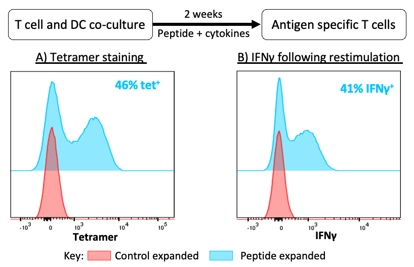
Figure 8: Antigen specific T cell expansion in an autologous moDC co-culture. Naïve T cells and CD14+ monocytes were isolated from healthy human blood samples. Following the differentiation of dendritic cells, naïve T cells were added for a two-week co-culture with tumor-associated antigen peptide and cytokine stimulation. Antigen specific T cell expansion was then assessed. A) T cells were stained with tetramer to detect antigen specific T cells by flow cytometry. B) CD8 T cells were restimulated with peptide alongside autologous CD14+ monocytes in the presence of Golgi plug for four hours, and IFNγ expression was then measured by flow cytometry.
Murine Functional Dendritic Cell Assays
Like humans, the frequencies of primary circulating dendritic cell in rodents are very low. Bone marrow-derived dendritic cell (BMDC) therefore provide a suitable source of dendritic cell for screening assays.
BMDC can be used in similar assays to those described for human cells. The use of TCR transgenic T cells provides the advantage of being able to study naïve T cell responses and to investigate how intervening in dendritic cell differentiation and polarization can alter T cell responses to cognate antigen.
The example data here shows a model utilizing OT-I CD8 TCR transgenic T cells, which are specific for the SIINFEKL peptide derived from ovalbumin. The ability of therapeutically treated BMDC that have been pulsed with SIINFEKL peptide or fed with whole OVA protein to drive antigen-specific proliferation, cytokine production, and effector function of OT-I CD8 T cells can then be assessed.
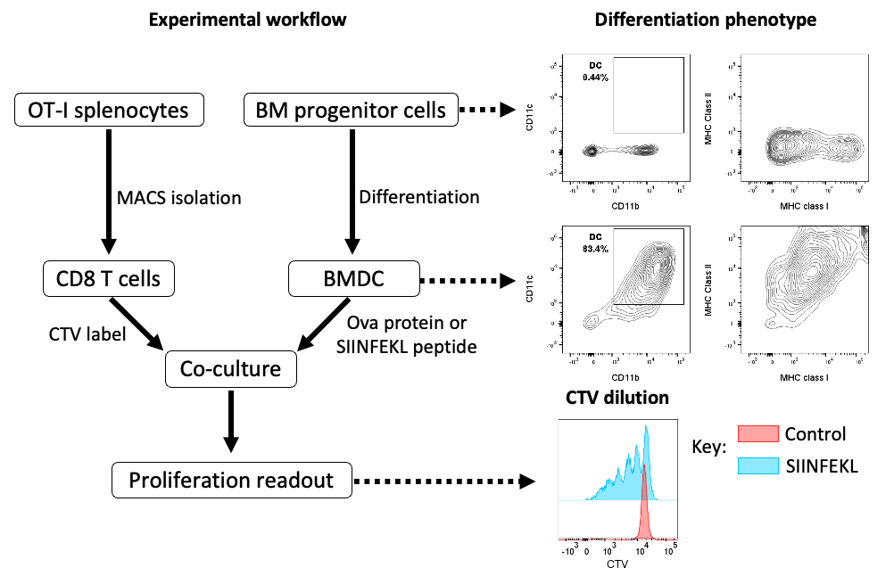
Figure 9: Murine bone marrow-derived dendritic cell and T cell coculture. Bone marrow (BM) progenitor cells were isolated from C57BL/6 mice and cultured in vitro with IL-4 and GM-CSF to generate BMDC. Representative data for measuring the differentiation of these cells by flow cytometry is shown. Splenocytes from OT-I TCR transgenic mice were isolated and CD8 T cells were enriched by magnetic selection. The T cells were then labelled with CTV dye and co-cultured with BMDC that had been pulsed with SIINFEKL. Dilution of CTV was used as a readout for CD8 T cell proliferation.
Dendritic cell are a critical player in the activation and polarization of T cell and other lymphocyte responses, as well as in the modulation of the tissue microenvironment. With these dendritic cell assays, which can be used to accurately measure and manipulate inflammatory or tolerogenic responses, our expertise provides dendritic cell assay platform for determining MOA or efficacy of dendritic cell targeting therapeutics.
Frequently Asked Questions (FAQs) About Dendritic Cell Assays
-
What is the role of dendritic cells?
Dendritic cells are a key intersection between the innate and adaptive immune system. Their phenotype and function are shaped by the microenvironment or context in which they were activated. Dendritic cell play a critical role in shaping the ensuing adaptive immune response by either enforcing tolerance or enhancing T cell responses, which are largely determined by the activation status of the Dendritic cell. Dendritic cell patrol peripheral sites, such as the skin or the intestines, and are continuously sampling their environment to process and present antigens via MHC molecules to lymphocytes. Dendritic cells also play a critical role in activating other lymphocyte subsets, including innate subsets such as NK cells, through the secretion of IL-12 and IL-15.
-
Can you work with murine dendritic cells as well as primary human cells?
Yes, we have experience translating many of our assays for in vitro dendritic cell assay work with both murine and human cells.
-
Are you able to perform these dendritic cell assays with particular Dendritic Cell subsets?
We can isolate particular dendritic cell subsets based on surface marker expression. However, the limited availability of differentiated dendritic cell subsets from blood often means it is more practical to use established methods for differentiating monocytes into dendritic cell and applying dendritic cell maturation or inhibitory stimulus to these cells to differentiate them into a given phenotype for the most appropriate functional dendritic cell assay.
-
What advantages over polyclonal stimulation of T cells do the dendritic cell assays offer?
A model using dendritic cell offers a more physiologically relevant system, allowing an assessment of both T cell function and the ability of the key immune priming cell to stimulate a T cell response. Numerous well-known receptor: ligand interactions, commonly targeted in drug discovery programs, will not occur without the presence of dendritic cell. The inclusion of dendritic cell assay also allows for antigen-specific responses to be assessed. This opens up the possibility to work with an antigen specific to the disease model, making it more translationally relevant, and to determine whether a novel peptide is immunogenic.
-
How can we use genetic modification of dendritic cell to validate our model?
Through lentiviral transduction or nucleofection we can introduce an antigen of interest, allowing for an assessment of physiologically relevant primary cell systems to screen for therapeutics that modulate antigen processing and presentation by the dendritic cell to T cells. Alternatively, we can introduce gene knockdown to validate pathway mechanisms within a dendritic cell assay.
-
Can you tailor these models to fit our requirements?
We are happy to discuss and customize any of our dendritic cell assays to meet the requirements of your project. Our expert immunologists can help by suggesting what parameters can be changed to tailor the setup and readouts of the dendritic cell assays to suit your needs.
-
How can I determine which therapeutic area we should target using dendritic cell assay?
This is obviously a complex question to answer, however as a starting point, a therapeutic that enhanced dendritic cell activation, and subsequently T cell function, might be targeted towards vaccinology or immune oncology. One that suppressed dendritic cell function and did not initiate T cell responses, or even inhibited existing T cell responses, may be more appropriately targeted towards autoimmune disease or inflammatory conditions.
Bieber, K. et al. Dendritic cell development in infection. Mol Immunol, 121, Pages 111-117 (2020)