
Orthotopic Tumor Model Studies
Orthotopic models involve the seeding of tumor cell lines or patient-derived cell xenografts into animal models. This strategy allows us to assess tumor development in a relevant environment and evaluate efficacy in a preclinical tumor model that mimics the disease process in humans. With orthotopic models, we can closely monitor and accurately quantify primary tumor growth, metastatic activity, and response to therapy scenarios.
Charles River offers orthotopic models across a full range of tumor histotypes to provide the most disease-relevant tumor environment in which to test your drug.
AACR Annual Meeting 2026
No matter the circumstances, cancer patients are waiting on new therapies. View our presentations and posters from the AACR Annual Meeting, and browse our resources so you can transform your oncology research with speed and simplicity.
Transform Your Research
We can evaluate disease progression through a variety of methods, including clinical signs, survival study design, and our imaging platform that has both in vivo and ex vivo capabilities:
- High-throughput or high-resolution imaging
- Real-time in-life fluorescence and bioluminescence imaging (IVIS)
- 3D optical tomographic reconstructions for both bioluminescence and fluorescence
- µCT X-ray for anatomical reference of signal
With human disease-specific tumor microenvironments and in-life imaging, Charles River’s orthotopic models are ideal for elucidating mode of action, monitoring patient-relevant responses, and targeting metastatic mechanisms.
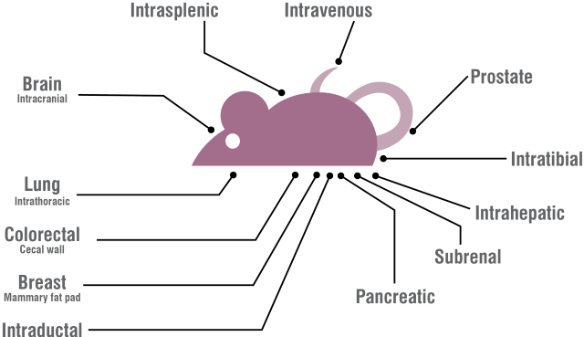
Figure 1: Diagram of Orthotopic Tumor Model
Oncology Preclinical Study Planning Toolkit
Take control of your efficacy study by getting instant access to study pricing, cancer model data, and connection to our oncology specialists, so that you feel supported on your oncology efficacy journey.
Find the Right Model
Charles River's Orthotopic Models
-
Ascites Model of Solid Cancer
Different cancer cell lines, such as ovarian cancer cell line A2780-luc or OVXF 1023-iRFP, are injected intraperitoneally to mimic the clinical symptoms of cancer-induced ascites. Tumor load is monitored by optical imaging.
Available Cell Lines
- Human - OVF1023
-
Bladder Model of Cancer
Bladder cancer cell lines, such as MBT2 iRFP, are directly implanted into the bladder layer in C3H/He mice. Effect of test agents is evaluated using optical imaging
Available Cell Lines
- Murine - MBT2
-
Breast Cancer Models
Cancer lines such as BT474 can be injected directly into the mammary fat pad of the mouse in this orthotopic tumor model of cancer.
Available Cell Lines
- Human - MCF-7, SK-BR-3, MDA-MB231,
- Murine - 4T1-luc
-
Colon Cancer Models
Available Cell Lines
- Human - 269, 1103, Caco-2, COLO 205, Difi, DLD-1, HCC-2998, HCT 116, HT-29, KM12, KM20L2, LS-174T, RKO, SW480, SW620
- Murine - CT26, MC38
-
Intracranial Orthotopic Model
Intracranial injection of tumor cells, such as CMV U87MG human glioma cells, mimics tumor growth in the brain environment. Test agent effects are evaluated based on optical imaging and scoring of neurological observations.
Available Cell Lines
- Human - U87MG
Example data: Intracranial U87MG orthotopic model
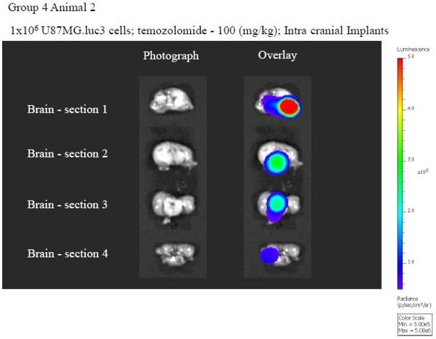
Intracranial U87MG tumor model, with ex vivo intracranial imaging at Day 65.
-
Intrasplenic Hepatic Colonization Model
Orthotopic seeding of human tumor cell lines, such as HT29 and HT29-luc2, allows assessment of tumor development in a hepatic environment and provides efficacy evaluation in a preclinical tumor model mimicking the disease process in humans. This allows for a more representative picture of tumor growth and response to therapy scenario to be realized.
Available Cell Lines
- Human - SK-HEP-1
-
Lung Cancer Models
Lung cancer cell lines, such as SHP-77, and NCI-H292 iRFP, are directly implanted into the lung tissue. Effect of test agents is evaluated using optical imaging.
Available Cell Lines - Small-Cell Lung Cancer (SCLC)
- Human - SHP-77
Available Cell Lines - Non-Small-Cell Lung Cancer (NSCLC)
- Human - H292
-
Metastatic Model of Solid Cancer
Different cancer cell lines, such as B16F10, from solid cancer are injected intravenously to mimic the metastatic process. Tumor load is monitored by optical imaging and/or flow cytometry on peripheral blood draws.
-
Ovarian Cancer Models
Ovarian cancer cell lines (e.g., A2780-luc) are implanted into the mouse models.
Available Cell Lines
- Human - OVCAR-3, A2780, OVXF 1023, SKOV-3
-
Pancreatic Models
Pancreatic tumor cell lines (e.g., MiaPaCa-2, Panc02) are implanted into the mouse pancreas.
Available Cell Lines
- Human - Panc02, MiaPaCa-2
-
Prostate Orthotopic Models
Prostate cancer cell lines (e.g., PC3, LNCAP, DU145) are implanted into the prostate tissue of the mouse model.
Available Cell Lines
- Human - PC-3, PC3M, DU145

Orthotopic Models in Prostate Cancer Research
Learn more about the establishment of clinically relevant luciferase labeled prostate cancer xenograft models: metastatic PC3M and non-metastatic DU145 cell lines, to test existing and novel therapies.
Download the Poster -
Renal Sub-Capsule Orthotopic Model of Cancer
The renal orthotopic tumor model was developed for the efficacy evaluation of novel anticancer compounds using a tumor growth delay model that mimics the disease process in humans. Cell lines used in this tumor model include 786-O.
-
Skin Cancer Models
Available Cell Lines - Melanoma
- Human - A375
- Murine - B16F10
-
Systemic Leukemia/Lymphoma/Multiple Myeloma Tumor Models
In this disseminated orthotopic tumor model, hematological cell lines are injected, such as Raji-B, Granta-519, or Ramos, either intravenously, intrasplenic, or intratibial. Tumor load is monitored by optical imaging and flow cytometry on peripheral blood and clinical observations.
Available Cell Lines - Leukemia
- Human - ARH-77, CCRF-CEM, HEL92.1.7, HL-60, K562-CD19, Kasumi-1, KG-1, KG-1a, MEC-1, MEG-01, MOLM-13, MOLT-4, MV4-11, NALM-6, NOMO-1, OCI-AML2, OCI-AML3, RS4;11, THP-1
Available Cell Lines - Lymphoma
- Human - Granta-519, JURKAT, K-562, Mino, Namalwa, RajiB, RL, U937
- Murine - A20, L1210
Available Cell Lines - Multiple Myeloma
- Human - H929, L-363, MM.1R, MM.1S, RPMI-8226, U266

Patient-Derived Xenografts - Cancer Model Database
Support your in vitro, in vivo, and ex vivo studies with; user friendly search, new model data (including HLA typing, growth curves, and tumor images) and multi-parameter search options for all tumor model types (PDX and CDX)
Visit our Database
Combining our range of tumor models with quantifiable imaging data creates a truly translational platform in which to test your compounds. Charles River's expanding collection of luciferase transduced tumor cell lines and orthotopic Imaging models are key tools for evaluating the activity of cellular therapies in hematological and solid tumors.
Frequently Asked Questions (FAQs) About Orthotopic Models
-
What are orthotopic models?
Orthotopic models involve implantation of tumor cell lines or patient-derived cell xenografts into animal tumor models into the organ or tissue which matches the tumor histotype. This creates a more disease-relevant environment for the assessment of tumor growth, which can be analyzed by optical imaging.
-
How can I use imaging and orthotopic models to increase predictive and translational value?
Many tumor models are subcutaneous implants; however, these are less representative of human tumors as they aren’t situated in a relevant location or tumor microenvironment (TME). Implanted in the organ that matches the tumor histotype, orthotopic models therefore have a more relevant TME. Using imaging to track tumor growth in this context improves translation.
-
What are the advantages of orthoptic models?
Orthotopic models offer an organ- or tissue-specific tumor environment. When combining this disease-relevant environment with optical imaging, we can easily track and quantify tumor progression and metastasis in a way that is truly translational to the human disease state. Moreover, orthotopic models are cost-efficient, offering more rapid and predictable tumor growth than genetically engineered models.